• 文献检索
  • 文档翻译
  • 深度研究
  • 学术资讯
  • Suppr Zotero 插件Zotero 插件
  • 邀请有礼
  • 套餐&价格
  • 历史记录
应用&插件
Suppr Zotero 插件Zotero 插件浏览器插件Mac 客户端Windows 客户端微信小程序
定价
高级版会员购买积分包购买API积分包
服务
文献检索文档翻译深度研究API 文档MCP 服务
关于我们
关于 Suppr公司介绍联系我们用户协议隐私条款
关注我们

Suppr 超能文献

核心技术专利:CN118964589B侵权必究
粤ICP备2023148730 号-1Suppr @ 2026

文献检索

告别复杂PubMed语法,用中文像聊天一样搜索,搜遍4000万医学文献。AI智能推荐,让科研检索更轻松。

立即免费搜索

文件翻译

保留排版,准确专业,支持PDF/Word/PPT等文件格式,支持 12+语言互译。

免费翻译文档

深度研究

AI帮你快速写综述,25分钟生成高质量综述,智能提取关键信息,辅助科研写作。

立即免费体验

减压对大鼠下颌骨骨髓炎骨愈合的影响。

Decompression effects on bone healing in rat mandible osteomyelitis.

机构信息

Department of Oral and Maxillofacial Surgery, Dental Research Institute, School of Dentistry, Seoul National University, 101 Daehak-ro, Jongno-gu, Seoul, 03080, Korea.

出版信息

Sci Rep. 2021 Jun 3;11(1):11673. doi: 10.1038/s41598-021-91104-7.

DOI:10.1038/s41598-021-91104-7
PMID:34083570
原文链接:https://pmc.ncbi.nlm.nih.gov/articles/PMC8175588/
Abstract

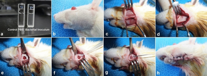
Osteomyelitis (OM) of the jaw is usually caused by a chronic odontogenic infection. Decompression is the release the intraluminal pressure in the cystic cavity allowing gradual bone growth from the periphery. The aim of this study was to analyze the effectiveness of decompression in an OM jaw model. A 4-mm-diameter defect was made on mandibles of fourteen Sprague-Dawley rats and inoculated with S. aureus (20 μl of 1 × 10 CFU/ml) injection. Two weeks later, four groups were made as non-treatment (C1), only curettage (C2), curettage and decompression (E1), and curettage and decompression with normal saline irrigation (E2). After four weeks, each group was analyzed. Most micro-CT parameters, including bone mineral density [0.87 (± 0.08) g/cm] with bone volume [0.73 (± 0.08) mm] was higher in E2 group than that of C1 group (p = 0.04, p = 0.05, respectively). E2 group in histology showed the highest number of osteocytes than those of control groups, 91.00 (± 9.90) (p = 0.002). OPN were expressed strongly in the E1 ("5": 76-100%) that those of other groups. Decompression drains induced advanced bone healing compared to that of curettage alone. Therefore, it could be recommended to use decompressive drain for enhancing the jaw OM management.

摘要

颌骨骨髓炎(OM)通常由慢性牙源性感染引起。减压是释放囊腔腔内的压力,允许骨从周围逐渐生长。本研究旨在分析颌骨 OM 模型中减压的效果。在 14 只 Sprague-Dawley 大鼠的下颌骨上制造一个 4 毫米直径的缺损,并接种金黄色葡萄球菌(20 μl 1×10 CFU/ml)注射。两周后,将四个组分为未治疗(C1)、仅刮除(C2)、刮除和减压(E1)以及刮除和减压加生理盐水冲洗(E2)。四周后,对每组进行分析。大多数微 CT 参数,包括骨矿物质密度[0.87(±0.08)g/cm]和骨体积[0.73(±0.08)mm]在 E2 组均高于 C1 组(p=0.04,p=0.05)。组织学上,E2 组的骨细胞数量最高,为 91.00(±9.90)(p=0.002)。E1 组的 OPN 表达较强(“5”:76-100%),高于其他组。减压引流器可诱导颌骨 OM 管理中比单独刮除术更先进的骨愈合。因此,建议使用减压引流器来增强颌骨 OM 的管理。

https://cdn.ncbi.nlm.nih.gov/pmc/blobs/387d/8175588/398bd4a3b0c4/41598_2021_91104_Fig8_HTML.jpg
https://cdn.ncbi.nlm.nih.gov/pmc/blobs/387d/8175588/a07f7e83c67e/41598_2021_91104_Fig1_HTML.jpg
https://cdn.ncbi.nlm.nih.gov/pmc/blobs/387d/8175588/0e56d75000ae/41598_2021_91104_Fig2_HTML.jpg
https://cdn.ncbi.nlm.nih.gov/pmc/blobs/387d/8175588/335e5542642e/41598_2021_91104_Fig3_HTML.jpg
https://cdn.ncbi.nlm.nih.gov/pmc/blobs/387d/8175588/9471d921f32b/41598_2021_91104_Fig4_HTML.jpg
https://cdn.ncbi.nlm.nih.gov/pmc/blobs/387d/8175588/a2d72a72ff65/41598_2021_91104_Fig5_HTML.jpg
https://cdn.ncbi.nlm.nih.gov/pmc/blobs/387d/8175588/ded73914bc68/41598_2021_91104_Fig6_HTML.jpg
https://cdn.ncbi.nlm.nih.gov/pmc/blobs/387d/8175588/6dc19765ea41/41598_2021_91104_Fig7_HTML.jpg
https://cdn.ncbi.nlm.nih.gov/pmc/blobs/387d/8175588/398bd4a3b0c4/41598_2021_91104_Fig8_HTML.jpg

相似文献

1
Decompression effects on bone healing in rat mandible osteomyelitis.减压对大鼠下颌骨骨髓炎骨愈合的影响。
Sci Rep. 2021 Jun 3;11(1):11673. doi: 10.1038/s41598-021-91104-7.
2
Decompressive effects of draining tube on suppurative and sclerosing osteomyelitis in the jaw.排脓管对颌骨化脓性硬化性骨髓炎的减压作用。
BMC Musculoskelet Disord. 2021 May 22;22(1):469. doi: 10.1186/s12891-021-04340-3.
3
Vancomycin-laden calcium phosphate-calcium sulfate composite allows bone formation in a rat infection model.万古霉素负载的磷酸钙-硫酸钙复合材料可在大鼠感染模型中促进骨形成。
PLoS One. 2019 Sep 19;14(9):e0222034. doi: 10.1371/journal.pone.0222034. eCollection 2019.
4
Effect of bisphosphonates on healing of tooth extraction wounds in infectious osteomyelitis of the jaw.双膦酸盐对颌骨感染性骨髓炎拔牙创愈合的影响。
Bone. 2021 Feb;143:115611. doi: 10.1016/j.bone.2020.115611. Epub 2020 Aug 21.
5
Bisphosphonates inhibit bone remodeling in the jaw bones of rats and delay healing following tooth extractions.双膦酸盐会抑制大鼠颌骨的骨重塑,并延迟拔牙后的愈合。
Oral Oncol. 2014 May;50(5):485-90. doi: 10.1016/j.oraloncology.2014.02.013. Epub 2014 Mar 12.
6
Effects of pentoxifylline and tocopherol on a rat-irradiated jaw model using micro-CT cortical bone analysis.使用微 CT 皮质骨分析研究己酮可可碱和生育酚对大鼠照射下颌模型的影响。
Eur Arch Otorhinolaryngol. 2019 Dec;276(12):3443-3452. doi: 10.1007/s00405-019-05600-8. Epub 2019 Aug 14.
7
Evaluation of single-dose applied teriparatide effect on bone healing with histomorphometric and micro-ct analysis.单次应用特立帕肽对骨愈合的影响的评估:组织形态计量学和 micro-CT 分析。
J Craniomaxillofac Surg. 2021 Feb;49(2):98-103. doi: 10.1016/j.jcms.2020.12.004. Epub 2020 Dec 17.
8
Low-dose infectivity of Staphylococcus aureus (SMH strain) in traumatized rat tibiae provides a model for studying early events in contaminated bone injuries.金黄色葡萄球菌(SMH菌株)在创伤大鼠胫骨中的低剂量感染性为研究污染性骨损伤的早期事件提供了一个模型。
Comp Med. 2005 Apr;55(2):123-8.
9
Development of bone regeneration strategies using human periosteum-derived osteoblasts and oxygen-releasing microparticles in mandibular osteomyelitis model of miniature pig.利用人骨膜源性成骨细胞和释氧微球在小型猪下颌骨骨髓炎模型中开发骨再生策略。
J Biomed Mater Res A. 2019 Oct;107(10):2183-2194. doi: 10.1002/jbm.a.36728. Epub 2019 Jun 6.
10
Development of a fracture osteomyelitis model in the rat femur.建立大鼠股骨骨折并骨髓炎模型。
J Orthop Res. 2011 Jan;29(1):131-7. doi: 10.1002/jor.21188.

引用本文的文献

1
Electron-donable heterojunctions with synergetic Ru-Cu pair sites for biocatalytic microenvironment modulations in inflammatory mandible defects.具有协同 Ru-Cu 对位点的给电子型异质结,用于炎症性下颌骨缺损中的生物催化微环境调控。
Nat Commun. 2024 Nov 6;15(1):9592. doi: 10.1038/s41467-024-53824-y.
2
Choosing the right animal model for osteomyelitis research: Considerations and challenges.选择合适的骨髓炎研究动物模型:考量因素与挑战。
J Orthop Translat. 2023 Nov 29;43:47-65. doi: 10.1016/j.jot.2023.10.001. eCollection 2023 Nov.
3
In vitro and in vivo assessment of caprine origin Staphylococcus aureus ST398 strain UTCVM1 as an osteomyelitis pathogen.

本文引用的文献

1
Decompressive effects of draining tube on suppurative and sclerosing osteomyelitis in the jaw.排脓管对颌骨化脓性硬化性骨髓炎的减压作用。
BMC Musculoskelet Disord. 2021 May 22;22(1):469. doi: 10.1186/s12891-021-04340-3.
2
Is There a Governing Role of Osteocytes in Bone Tissue Regeneration?成骨细胞在骨组织再生中是否具有支配作用?
Curr Osteoporos Rep. 2020 Oct;18(5):541-550. doi: 10.1007/s11914-020-00610-6.
3
The ARRIVE guidelines 2.0: Updated guidelines for reporting animal research.ARRIVE 指南 2.0:报告动物研究的更新指南。
山羊源金黄色葡萄球菌 ST398 株 UTCVM1 作为骨髓炎病原体的体外和体内评估。
Front Cell Infect Microbiol. 2022 Nov 24;12:1015655. doi: 10.3389/fcimb.2022.1015655. eCollection 2022.
4
Osteomyelitis of the Jaw in COVID-19 Patients: A Rare Condition With a High Risk for Severe Complications.新冠肺炎患者颌骨骨髓炎:一种罕见但严重并发症风险高的病症
Front Surg. 2022 Jul 1;9:867088. doi: 10.3389/fsurg.2022.867088. eCollection 2022.
5
Histologic features of bone regenerated by means of negative pressure in the context of odontogenic keratocyst.在牙源性角化囊肿的背景下,负压再生的骨组织学特征。
Oral Maxillofac Surg. 2023 Sep;27(3):421-426. doi: 10.1007/s10006-022-01080-z. Epub 2022 May 28.
6
Role of Animal Models to Advance Research of Bacterial Osteomyelitis.动物模型在推进细菌性骨髓炎研究中的作用
Front Vet Sci. 2022 Apr 26;9:879630. doi: 10.3389/fvets.2022.879630. eCollection 2022.
PLoS Biol. 2020 Jul 14;18(7):e3000410. doi: 10.1371/journal.pbio.3000410. eCollection 2020 Jul.
4
Bone Regeneration Potential of Uncalcined and Unsintered Hydroxyapatite/Poly l-lactide Bioactive/Osteoconductive Sheet Used for Maxillofacial Reconstructive Surgery: An In Vivo Study.用于颌面重建手术的未煅烧和未烧结羟基磷灰石/聚左旋乳酸生物活性/骨传导片的骨再生潜力:一项体内研究。
Materials (Basel). 2019 Sep 11;12(18):2931. doi: 10.3390/ma12182931.
5
Chronic non-bacterial osteomyelitis in the jaw.颌骨慢性非细菌性骨髓炎
J Korean Assoc Oral Maxillofac Surg. 2019 Apr;45(2):68-75. doi: 10.5125/jkaoms.2019.45.2.68. Epub 2019 Apr 29.
6
Therapeutic efficacy of vacuum sealing drainage-assisted irrigation in patients with severe multiple-space infections in the oral, maxillofacial, and cervical regions.负压封闭引流辅助灌洗治疗口腔颌颈部多间隙重度感染的疗效。
J Craniomaxillofac Surg. 2019 May;47(5):837-841. doi: 10.1016/j.jcms.2019.01.031. Epub 2019 Feb 1.
7
Quantification of the mandibular defect healing by micro-CT morphometric analysis in rats.利用微 CT 形态计量分析法对大鼠下颌骨缺损愈合的定量分析。
J Craniomaxillofac Surg. 2018 Dec;46(12):2203-2213. doi: 10.1016/j.jcms.2018.09.022. Epub 2018 Sep 25.
8
Distraction Sugosteogenesis: Its Biologic Bases and Therapeutic Principles.牵引成骨术:其生物学基础与治疗原则
J Craniofac Surg. 2018 Nov;29(8):2088-2095. doi: 10.1097/SCS.0000000000004892.
9
Principles and approaches for reproducible scoring of tissue stains in research.研究中组织染色可重复性评分的原则和方法。
Lab Invest. 2018 Jul;98(7):844-855. doi: 10.1038/s41374-018-0057-0. Epub 2018 May 30.
10
Immunoprecipitation high performance liquid chromatographic analysis of healing process in chronic suppurative osteomyelitis of the jaw.免疫沉淀高效液相色谱分析颌骨慢性化脓性骨髓炎愈合过程。
J Craniomaxillofac Surg. 2018 Jan;46(1):119-127. doi: 10.1016/j.jcms.2017.10.017. Epub 2017 Oct 27.