Columbia Center for Translational Immunology, Columbia University Medical Center, New York, NY, USA.
Perlmutter Cancer Center, New York University School of Medicine, New York, NY, USA.
Commun Biol. 2021 Jun 3;4(1):672. doi: 10.1038/s42003-021-02225-8.
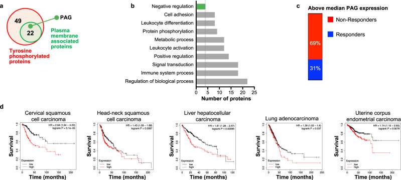
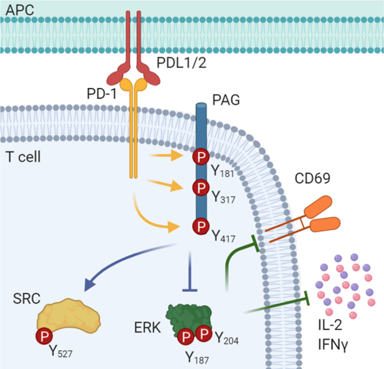
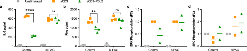
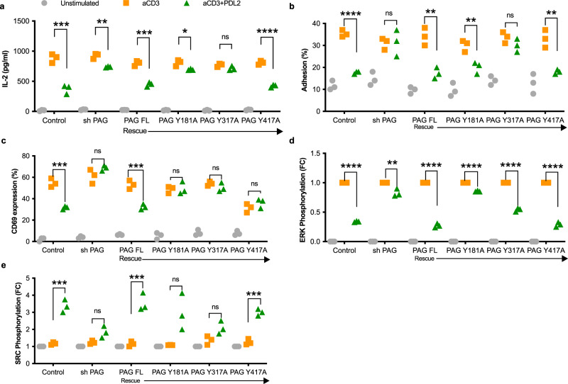
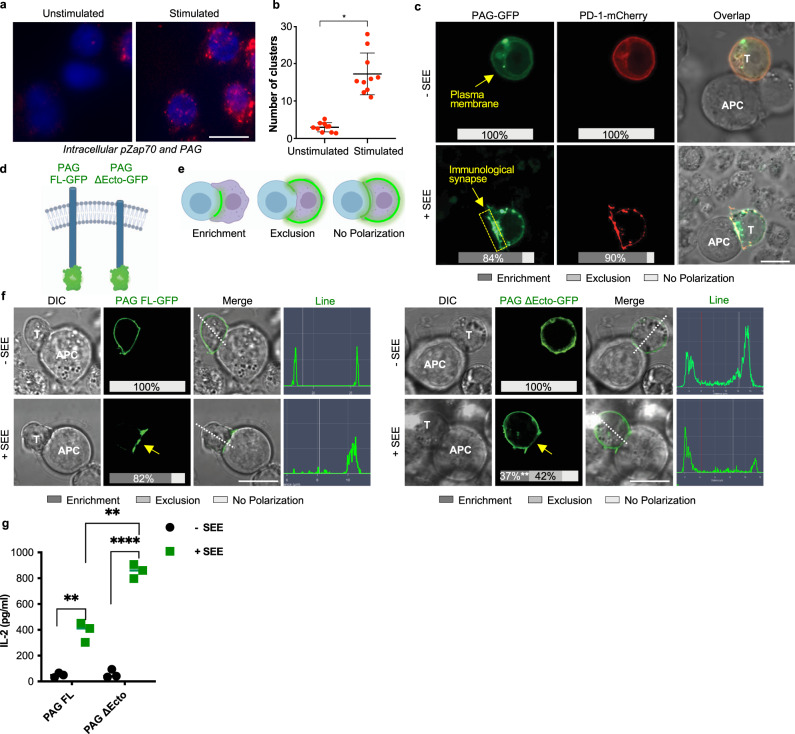
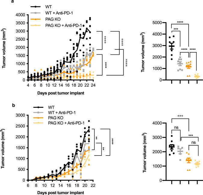
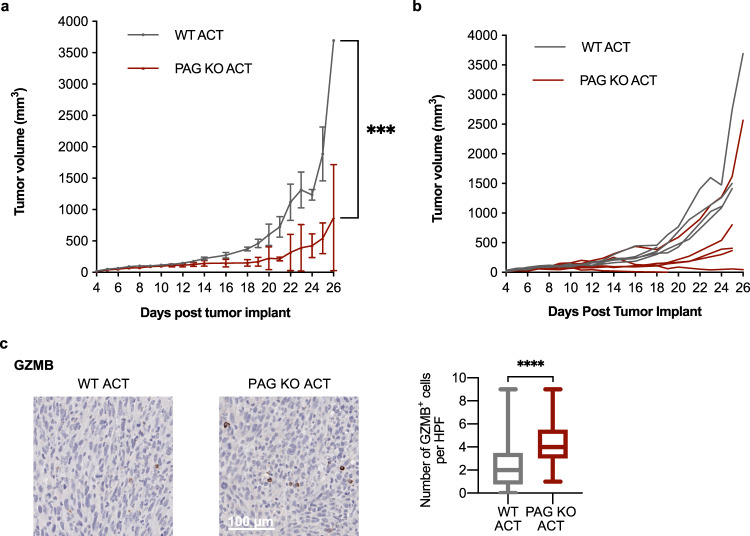
The inhibitory receptor PD-1 is expressed on T cells to inhibit select functions when ligated. The complete signaling mechanism downstream of PD-1 has yet to be uncovered. Here, we discovered phosphoprotein associated with glycosphingolipid-enriched microdomains 1 (PAG) is phosphorylated following PD-1 ligation and associate this with inhibitory T cell function. Clinical cohort analysis correlates low PAG expression with increased survival from numerous tumor types. PAG knockdown in T cells prevents PD-1-mediated inhibition of cytokine secretion, cell adhesion, CD69 expression, and ERK phosphorylation, and enhances phosphorylation of SRC following PD-1 ligation. PAG overexpression rescues these effects. In vivo, PAG contributes greatly to the growth of two murine tumors, MC38 and B16, and limits T cell presence within the tumor. Moreover, PAG deletion sensitizes tumors to PD-1 blockade. Here PAG is established as a critical mediator of PD-1 signaling and as a potential target to enhance T cell activation in tumors.
抑制性受体 PD-1 在与配体结合时表达于 T 细胞上,以抑制特定功能。PD-1 下游的完整信号机制尚未被揭示。在这里,我们发现与富含神经酰胺的糖脂富集微区的磷酸化蛋白 1(PAG)在 PD-1 结合后发生磷酸化,并将其与抑制性 T 细胞功能相关联。临床队列分析表明,PAG 低表达与多种肿瘤类型的生存率提高相关。T 细胞中 PAG 的敲低可阻止 PD-1 介导的细胞因子分泌、细胞黏附、CD69 表达和 ERK 磷酸化的抑制,并增强 PD-1 结合后的 SRC 磷酸化。PAG 的过表达可挽救这些效应。在体内,PAG 极大地促进了 MC38 和 B16 两种鼠肿瘤的生长,并限制了肿瘤内 T 细胞的存在。此外,PAG 的缺失使肿瘤对 PD-1 阻断更为敏感。在这里,PAG 被确立为 PD-1 信号的关键介质,并可能成为增强肿瘤中 T 细胞激活的潜在靶点。