• 文献检索
  • 文档翻译
  • 深度研究
  • 学术资讯
  • Suppr Zotero 插件Zotero 插件
  • 邀请有礼
  • 套餐&价格
  • 历史记录
应用&插件
Suppr Zotero 插件Zotero 插件浏览器插件Mac 客户端Windows 客户端微信小程序
定价
高级版会员购买积分包购买API积分包
服务
文献检索文档翻译深度研究API 文档MCP 服务
关于我们
关于 Suppr公司介绍联系我们用户协议隐私条款
关注我们

Suppr 超能文献

核心技术专利:CN118964589B侵权必究
粤ICP备2023148730 号-1Suppr @ 2026

文献检索

告别复杂PubMed语法,用中文像聊天一样搜索,搜遍4000万医学文献。AI智能推荐,让科研检索更轻松。

立即免费搜索

文件翻译

保留排版,准确专业,支持PDF/Word/PPT等文件格式,支持 12+语言互译。

免费翻译文档

深度研究

AI帮你快速写综述,25分钟生成高质量综述,智能提取关键信息,辅助科研写作。

立即免费体验

早期胃肠道肿瘤光学诊断的系统评价

Systematic Review on Optical Diagnosis of Early Gastrointestinal Neoplasia.

作者信息

Wagner Andrej, Zandanell Stephan, Kiesslich Tobias, Neureiter Daniel, Klieser Eckhard, Holzinger Josef, Berr Frieder

机构信息

Department of Internal Medicine I, University Clinics Salzburg, Paracelsus Medical University, Müllner Hauptstrasse 48, 5020 Salzburg, Austria.

Laboratory for Tumour Biology and Experimental Therapies (TREAT), Center for Physiology, Pathophysiology and Biophysics-Salzburg and Nuremberg, Institute for Physiology and Pathophysiology-Salzburg, Paracelsus Medical University, 5020 Salzburg, Austria.

出版信息

J Clin Med. 2021 Jun 25;10(13):2794. doi: 10.3390/jcm10132794.

DOI:10.3390/jcm10132794
PMID:34202001
原文链接:https://pmc.ncbi.nlm.nih.gov/articles/PMC8269336/
Abstract

BACKGROUND

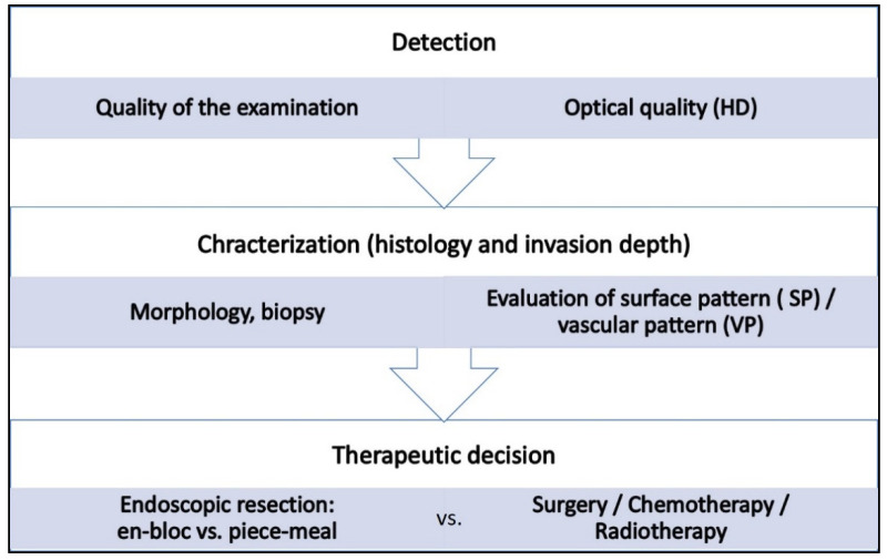
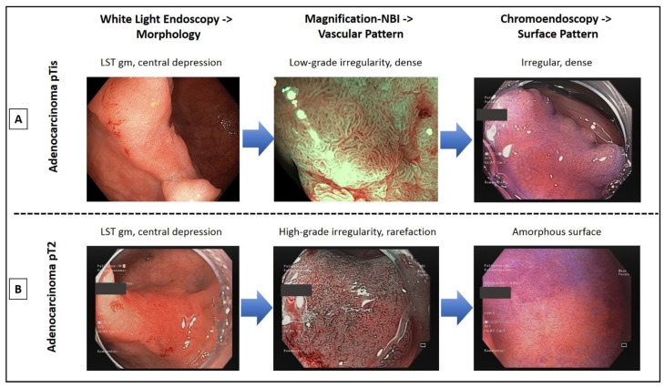
Meticulous endoscopic characterization of gastrointestinal neoplasias (GN) is crucial to the clinical outcome. Hereby the indication and type of resection (endoscopically, en-bloc or piece-meal, or surgical resection) are determined. By means of established image-enhanced (IEE) and magnification endoscopy (ME) GN can be characterized in terms of malignancy and invasion depth. In this context, the statistical evidence and accuracy of these diagnostic procedures should be elucidated. Here, we present a systematic review of the literature.

RESULTS

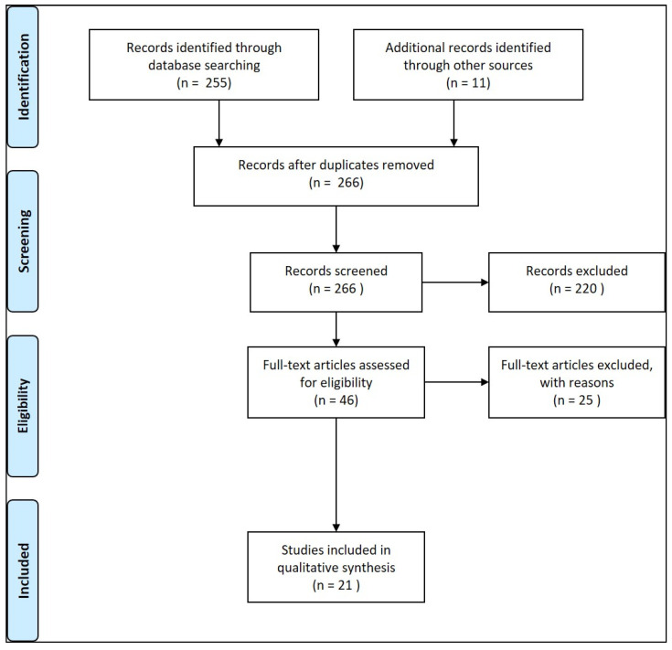
21 Studies could be found which met the inclusion criteria. In clinical prospective trials and meta-analyses, the diagnostic accuracy of >90% for characterization of malignant neoplasms could be documented, if ME with IEE was used in squamous cell esophageal cancer, stomach, or colonic GN.

CONCLUSIONS

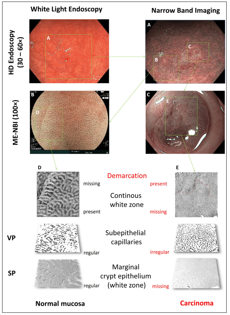
Currently, by means of optical diagnosis, today's gastrointestinal endoscopy is capable of determining the histological subtype, exact lateral spread, and depth of invasion of a lesion. The prerequisites for this are an exact knowledge of the anatomical structures, the endoscopic classifications based on them, and a systematic learning process, which can be supported by training courses. More prospective clinical studies are required, especially in the field of Barrett's esophagus and duodenal neoplasia.

摘要

背景

对胃肠道肿瘤(GN)进行细致的内镜特征描述对临床结果至关重要。据此确定切除的指征和类型(内镜下切除、整块切除或分片切除,或手术切除)。借助已确立的图像增强(IEE)和放大内镜(ME),可以从恶性程度和浸润深度方面对GN进行特征描述。在此背景下,应阐明这些诊断程序的统计学证据和准确性。在此,我们对文献进行了系统综述。

结果

发现有21项研究符合纳入标准。在临床前瞻性试验和荟萃分析中,如果在鳞状细胞食管癌、胃癌或结肠GN中使用ME联合IEE,对恶性肿瘤特征描述的诊断准确率可达90%以上。

结论

目前,通过光学诊断,当今的胃肠内镜能够确定病变的组织学亚型、确切的侧向扩散和浸润深度。做到这一点的前提是准确了解解剖结构、基于此的内镜分类以及系统的学习过程,培训课程可以对此提供支持。需要更多的前瞻性临床研究,尤其是在巴雷特食管和十二指肠肿瘤领域。

https://cdn.ncbi.nlm.nih.gov/pmc/blobs/c7a8/8269336/3a108b064e08/jcm-10-02794-g005.jpg
https://cdn.ncbi.nlm.nih.gov/pmc/blobs/c7a8/8269336/c526ce636cce/jcm-10-02794-g001.jpg
https://cdn.ncbi.nlm.nih.gov/pmc/blobs/c7a8/8269336/ce4c5dfa4fbf/jcm-10-02794-g002.jpg
https://cdn.ncbi.nlm.nih.gov/pmc/blobs/c7a8/8269336/10654e404d6d/jcm-10-02794-g003.jpg
https://cdn.ncbi.nlm.nih.gov/pmc/blobs/c7a8/8269336/5f844970584b/jcm-10-02794-g004.jpg
https://cdn.ncbi.nlm.nih.gov/pmc/blobs/c7a8/8269336/3a108b064e08/jcm-10-02794-g005.jpg

相似文献

1
Systematic Review on Optical Diagnosis of Early Gastrointestinal Neoplasia.早期胃肠道肿瘤光学诊断的系统评价
J Clin Med. 2021 Jun 25;10(13):2794. doi: 10.3390/jcm10132794.
2
Chromoendoscopy and magnification endoscopy for diagnosing esophageal cancer and dysplasia.用于诊断食管癌和发育异常的色素内镜检查及放大内镜检查
Thorac Surg Clin. 2004 Feb;14(1):87-94. doi: 10.1016/S1547-4127(04)00042-8.
3
Endoscopic Diagnosis of Superficial Gastrointestinal Cancer.内镜诊断消化道浅层癌。
Gastrointest Endosc Clin N Am. 2024 Jan;34(1):1-17. doi: 10.1016/j.giec.2023.08.003. Epub 2023 Sep 16.
4
Histological analysis of endoscopic resection specimens from 326 patients with Barrett's esophagus and early neoplasia.对326例巴雷特食管和早期肿瘤患者的内镜切除标本进行组织学分析。
Endoscopy. 2004 Sep;36(9):776-81. doi: 10.1055/s-2004-825802.
5
AGA Institute Clinical Practice Update: Endoscopic Submucosal Dissection in the United States.AGA 研究所临床实践更新:美国内镜黏膜下剥离术。
Clin Gastroenterol Hepatol. 2019 Jan;17(1):16-25.e1. doi: 10.1016/j.cgh.2018.07.041. Epub 2018 Aug 2.
6
Diagnosis and Management of Low-Grade Dysplasia in Barrett's Esophagus: Expert Review From the Clinical Practice Updates Committee of the American Gastroenterological Association.巴雷特食管低级别异型增生的诊断和管理:美国胃肠病学会临床实践更新委员会的专家综述。
Gastroenterology. 2016 Nov;151(5):822-835. doi: 10.1053/j.gastro.2016.09.040. Epub 2016 Oct 1.
7
Chromoendoscopy and magnification endoscopy in Barrett's esophagus.巴雷特食管的色素内镜检查和放大内镜检查
Gastrointest Endosc Clin N Am. 2003 Apr;13(2):269-77. doi: 10.1016/s1052-5157(03)00011-4.
8
Efficacy of radiofrequency ablation combined with endoscopic resection for barrett's esophagus with early neoplasia.射频消融联合内镜切除治疗 Barrett 食管伴早期肿瘤的疗效。
Clin Gastroenterol Hepatol. 2010 Jan;8(1):23-9. doi: 10.1016/j.cgh.2009.07.003. Epub 2009 Aug 11.
9
Endoscopic surveillance in Barrett's esophagus.巴雷特食管的内镜监测
Minerva Gastroenterol Dietol. 2002 Jun;48(2):63-71.
10
Endoscopic features of buried Barrett's mucosa.内镜下埋置型 Barrett 黏膜的特征。
Gastrointest Endosc. 2021 Jul;94(1):14-21. doi: 10.1016/j.gie.2020.12.031. Epub 2020 Dec 26.

引用本文的文献

1
A Rare Case of Small Bowel Ulceration Induced by COVID-19.一例由新冠病毒感染引起的小肠溃疡罕见病例。
J Inflamm Res. 2025 May 10;18:6123-6131. doi: 10.2147/JIR.S507209. eCollection 2025.
2
Assessing the effectiveness of texture and color enhancement imaging versus white-light endoscopy in detecting gastrointestinal lesions: A systematic review and meta-analysis.评估纹理和颜色增强成像与白光内镜检查在检测胃肠道病变中的有效性:一项系统评价和荟萃分析。
DEN Open. 2025 Apr 30;6(1):e70128. doi: 10.1002/deo2.70128. eCollection 2026 Apr.
3
Impact of narrow band imaging in prediction of histology of advanced colorectal neoplasia.

本文引用的文献

1
Curriculum for optical diagnosis training in Europe: European Society of Gastrointestinal Endoscopy (ESGE) Position Statement.欧洲光学诊断培训课程:欧洲胃肠道内窥镜学会(ESGE)立场声明。
Endoscopy. 2020 Oct;52(10):899-923. doi: 10.1055/a-1231-5123. Epub 2020 Sep 3.
2
Suboptimal endoscopic cancer recognition in colorectal lesions in a national bowel screening programme.在一项全国性肠道筛查计划中,结直肠病变的内镜下癌症识别欠佳。
Gut. 2020 Jun;69(6):977-980. doi: 10.1136/gutjnl-2018-316882. Epub 2019 Dec 10.
3
Advanced imaging for detection and differentiation of colorectal neoplasia: European Society of Gastrointestinal Endoscopy (ESGE) Guideline - Update 2019.
窄带成像在预测晚期结直肠肿瘤组织学方面的影响。
Sci Rep. 2025 Jan 9;15(1):1414. doi: 10.1038/s41598-025-85669-w.
4
Optical imaging for screening and early cancer diagnosis in low-resource settings.资源匮乏地区用于癌症筛查和早期诊断的光学成像技术
Nat Rev Bioeng. 2024 Jan;2(1):25-43. doi: 10.1038/s44222-023-00135-4. Epub 2023 Dec 22.
5
The oncogenic mechanisms of the Janus kinase-signal transducer and activator of transcription pathway in digestive tract tumors.Janus激酶-信号转导与转录激活因子通路在消化道肿瘤中的致癌机制。
Cell Commun Signal. 2024 Jan 25;22(1):68. doi: 10.1186/s12964-023-01421-9.
6
Endoscopic diagnosis and treatment of superficial non-ampullary duodenal epithelial tumors: A review.浅表性非壶腹十二指肠上皮肿瘤的内镜诊断与治疗:综述
J Transl Int Med. 2023 Sep 2;11(3):206-215. doi: 10.2478/jtim-2023-0102. eCollection 2023 Sep.
7
New method for real-time visualization and quantitative characterization of early colorectal cancer in endoscopy: a pilot study.内镜检查中早期结直肠癌实时可视化与定量表征的新方法:一项初步研究。
Endosc Int Open. 2022 Aug 15;10(8):E1147-E1154. doi: 10.1055/a-1847-2820. eCollection 2022 Aug.
8
TXI (Texture and Color Enhancement Imaging) for Serrated Colorectal Lesions.用于锯齿状结直肠病变的TXI(纹理和颜色增强成像)
J Clin Med. 2021 Dec 27;11(1):119. doi: 10.3390/jcm11010119.
9
The Role of Diet and Lifestyle in Early-Onset Colorectal Cancer: A Systematic Review.饮食与生活方式在早发性结直肠癌中的作用:一项系统评价
Cancers (Basel). 2021 Nov 25;13(23):5933. doi: 10.3390/cancers13235933.
10
Can Ultrasound Elastography Discriminate between Rectal Adenoma and Cancer? A Systematic Review.超声弹性成像能否鉴别直肠腺瘤和癌症?一项系统评价。
Cancers (Basel). 2021 Aug 18;13(16):4158. doi: 10.3390/cancers13164158.
结直肠肿瘤的检测和鉴别诊断的高级影像学:欧洲胃肠道内镜学会(ESGE)指南-2019 年更新。
Endoscopy. 2019 Dec;51(12):1155-1179. doi: 10.1055/a-1031-7657. Epub 2019 Nov 11.
4
Forceps Biopsies Are Not Reliable in the Workup of Large Colorectal Lesions Referred for Endoscopic Resection: Should They Be Abandoned?对于因内镜切除而转诊的大型结直肠病变,活检钳活检不可靠:是否应放弃?
Dis Colon Rectum. 2019 Sep;62(9):1063-1070. doi: 10.1097/DCR.0000000000001440.
5
Narrow-Band Imaging for Detection of Neoplasia at Colonoscopy: A Meta-analysis of Data From Individual Patients in Randomized Controlled Trials.窄带成像在结肠镜检查中用于检测肿瘤:来自随机对照试验中个体患者数据的荟萃分析。
Gastroenterology. 2019 Aug;157(2):462-471. doi: 10.1053/j.gastro.2019.04.014. Epub 2019 Apr 15.
6
Objective validity of the Japan Narrow-Band Imaging Expert Team classification system for the differential diagnosis of colorectal polyps.日本窄带成像专家团队分类系统对结直肠息肉鉴别诊断的客观有效性。
Dig Endosc. 2019 Sep;31(5):544-551. doi: 10.1111/den.13393. Epub 2019 May 7.
7
Accuracy of the Narrow-Band Imaging International Colorectal Endoscopic Classification System in Identification of Deep Invasion in Colorectal Polyps.窄带成像国际结直肠内镜分类系统对结直肠息肉浸润深度的识别准确性。
Gastroenterology. 2019 Jan;156(1):75-87. doi: 10.1053/j.gastro.2018.10.004. Epub 2018 Oct 6.
8
Newly developed magnifying endoscopic classification of the Japan Esophageal Society to identify superficial Barrett's esophagus-related neoplasms.日本食管学会新开发的放大内镜分类法,用于识别浅表性巴雷特食管相关肿瘤。
Esophagus. 2018 Jun 19;15(3):153-9. doi: 10.1007/s10388-018-0623-y.
9
Diagnostic Accuracy of Magnifying Endoscopy with Narrow Band Imaging and Its Diagnostic Value for Invasion Depth Staging in Esophageal Squamous Cell Carcinoma: A Systematic Review and Meta-Analysis.放大内镜窄带成像技术对食管鳞癌浸润深度诊断准确性的系统评价和 Meta 分析。
Biomed Res Int. 2018 May 20;2018:8591387. doi: 10.1155/2018/8591387. eCollection 2018.
10
High-definition colonoscopy versus Endocuff versus EndoRings versus full-spectrum endoscopy for adenoma detection at colonoscopy: a multicenter randomized trial.高清结肠镜检查与 Endocuff 与 EndoRings 与全谱内镜检查在结肠镜检查中对腺瘤检测的比较:一项多中心随机试验。
Gastrointest Endosc. 2018 Aug;88(2):335-344.e2. doi: 10.1016/j.gie.2018.02.043. Epub 2018 Mar 9.