• 文献检索
  • 文档翻译
  • 深度研究
  • 学术资讯
  • Suppr Zotero 插件Zotero 插件
  • 邀请有礼
  • 套餐&价格
  • 历史记录
应用&插件
Suppr Zotero 插件Zotero 插件浏览器插件Mac 客户端Windows 客户端微信小程序
定价
高级版会员购买积分包购买API积分包
服务
文献检索文档翻译深度研究API 文档MCP 服务
关于我们
关于 Suppr公司介绍联系我们用户协议隐私条款
关注我们

Suppr 超能文献

核心技术专利:CN118964589B侵权必究
粤ICP备2023148730 号-1Suppr @ 2026

文献检索

告别复杂PubMed语法,用中文像聊天一样搜索,搜遍4000万医学文献。AI智能推荐,让科研检索更轻松。

立即免费搜索

文件翻译

保留排版,准确专业,支持PDF/Word/PPT等文件格式,支持 12+语言互译。

免费翻译文档

深度研究

AI帮你快速写综述,25分钟生成高质量综述,智能提取关键信息,辅助科研写作。

立即免费体验

BrcaSeg:一种用于组织量化和组织病理学图像基因组相关性的深度学习方法。

BrcaSeg: A Deep Learning Approach for Tissue Quantification and Genomic Correlations of Histopathological Images.

机构信息

Guangdong Provincial Key Laboratory of Medical Image Processing, School of Biomedical Engineering, Southern Medical University, Guangzhou 510515, China.

National-Regional Key Technology Engineering Laboratory for Medical Ultrasound, School of Biomedical Engineering, Health Science Center, Shenzhen University, Shenzhen 518060, China; Department of Medicine, Indiana University School of Medicine, Indianapolis, IN 46202, USA.

出版信息

Genomics Proteomics Bioinformatics. 2021 Dec;19(6):1032-1042. doi: 10.1016/j.gpb.2020.06.026. Epub 2021 Jul 17.

DOI:10.1016/j.gpb.2020.06.026
PMID:34280546
原文链接:https://pmc.ncbi.nlm.nih.gov/articles/PMC9403022/
Abstract

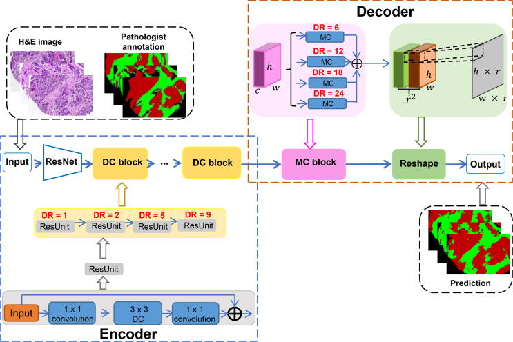
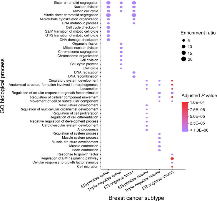
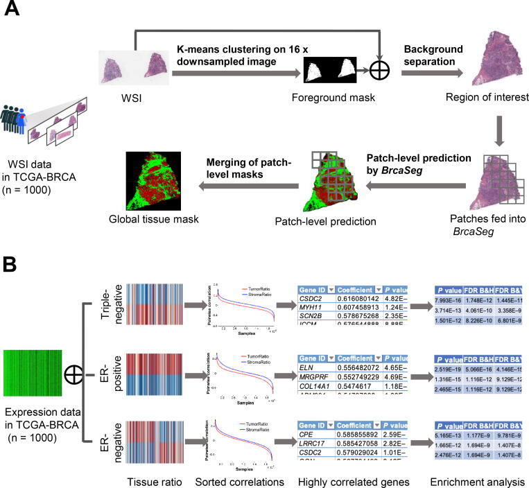
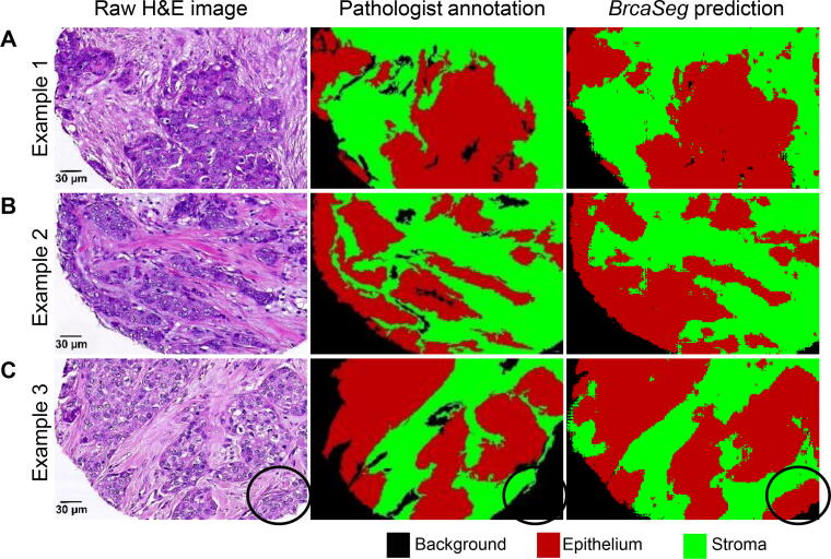
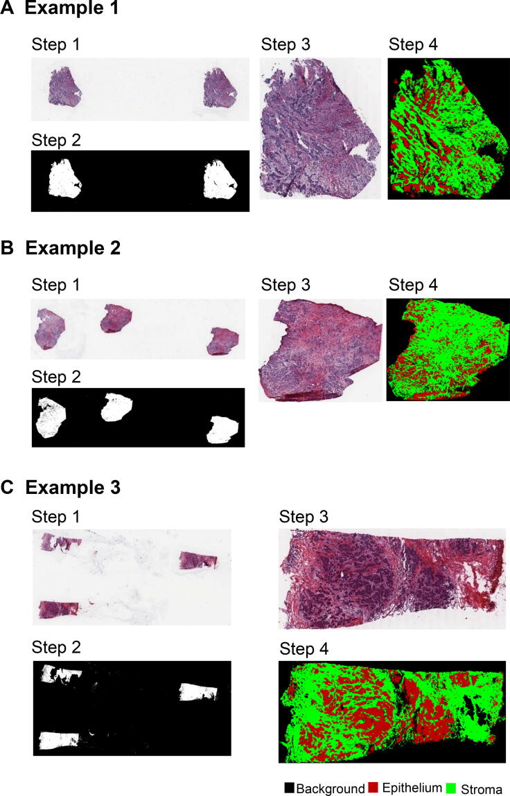
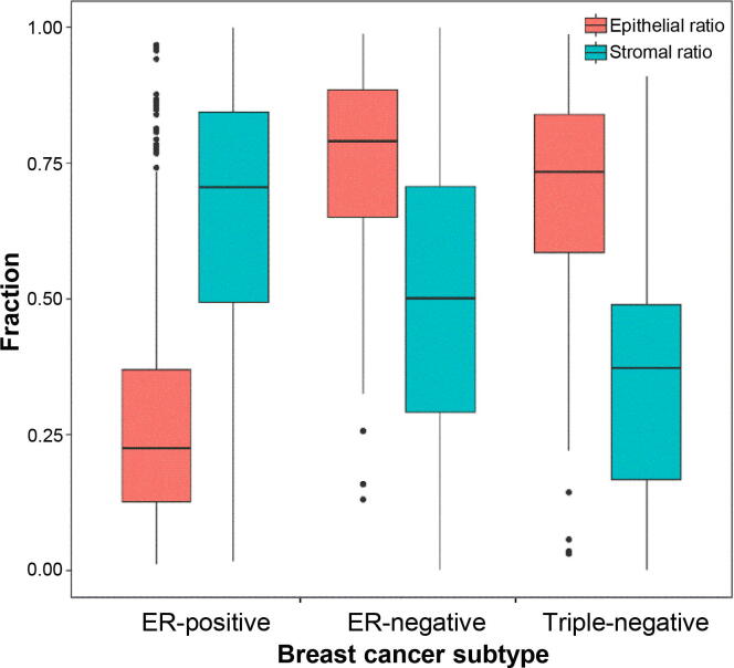
Epithelial and stromal tissues are components of the tumor microenvironment and play a major role in tumor initiation and progression. Distinguishing stroma from epithelial tissues is critically important for spatial characterization of the tumor microenvironment. Here, we propose BrcaSeg, an image analysis pipeline based on a convolutional neural network (CNN) model to classify epithelial and stromal regions in whole-slide hematoxylin and eosin (H&E) stained histopathological images. The CNN model is trained using well-annotated breast cancer tissue microarrays and validated with images from The Cancer Genome Atlas (TCGA) Program. BrcaSeg achieves a classification accuracy of 91.02%, which outperforms other state-of-the-art methods. Using this model, we generate pixel-level epithelial/stromal tissue maps for 1000 TCGA breast cancer slide images that are paired with gene expression data. We subsequently estimate the epithelial and stromal ratios and perform correlation analysis to model the relationship between gene expression and tissue ratios. Gene Ontology (GO) enrichment analyses of genes that are highly correlated with tissue ratios suggest that the same tissue is associated with similar biological processes in different breast cancer subtypes, whereas each subtype also has its own idiosyncratic biological processes governing the development of these tissues. Taken all together, our approach can lead to new insights in exploring relationships between image-based phenotypes and their underlying genomic events and biological processes for all types of solid tumors. BrcaSeg can be accessed at https://github.com/Serian1992/ImgBio.

摘要

上皮组织和基质组织是肿瘤微环境的组成部分,在肿瘤的发生和发展中起着重要作用。区分基质和上皮组织对于肿瘤微环境的空间特征具有重要意义。在这里,我们提出了 BrcaSeg,这是一种基于卷积神经网络(CNN)模型的图像分析管道,用于对全幻灯片苏木精和伊红(H&E)染色组织病理学图像中的上皮和基质区域进行分类。该 CNN 模型使用经过充分注释的乳腺癌组织微阵列进行训练,并使用癌症基因组图谱(TCGA)计划的图像进行验证。BrcaSeg 的分类准确率达到 91.02%,优于其他最先进的方法。使用这个模型,我们为 1000 张 TCGA 乳腺癌幻灯片图像生成了像素级别的上皮/基质组织图谱,这些图像与基因表达数据配对。随后,我们估计上皮和基质的比例,并进行相关性分析,以建立基因表达与组织比例之间的关系模型。与组织比例高度相关的基因的基因本体论(GO)富集分析表明,同一组织与不同乳腺癌亚型中的相似生物学过程相关,而每个亚型也有其自身特有的控制这些组织发育的生物学过程。总的来说,我们的方法可以为探索基于图像的表型与其潜在的基因组事件和生物学过程之间的关系提供新的见解,适用于所有类型的实体瘤。BrcaSeg 可在 https://github.com/Serian1992/ImgBio 上访问。

https://cdn.ncbi.nlm.nih.gov/pmc/blobs/c4b4/9403022/14858c84552e/gr6.jpg
https://cdn.ncbi.nlm.nih.gov/pmc/blobs/c4b4/9403022/398894c1977f/gr1.jpg
https://cdn.ncbi.nlm.nih.gov/pmc/blobs/c4b4/9403022/8ce2b5fea35d/gr2.jpg
https://cdn.ncbi.nlm.nih.gov/pmc/blobs/c4b4/9403022/9955ae23daf7/gr3.jpg
https://cdn.ncbi.nlm.nih.gov/pmc/blobs/c4b4/9403022/505edb130f9d/gr4.jpg
https://cdn.ncbi.nlm.nih.gov/pmc/blobs/c4b4/9403022/0c1212855bc5/gr5.jpg
https://cdn.ncbi.nlm.nih.gov/pmc/blobs/c4b4/9403022/14858c84552e/gr6.jpg

相似文献

1
BrcaSeg: A Deep Learning Approach for Tissue Quantification and Genomic Correlations of Histopathological Images.BrcaSeg:一种用于组织量化和组织病理学图像基因组相关性的深度学习方法。
Genomics Proteomics Bioinformatics. 2021 Dec;19(6):1032-1042. doi: 10.1016/j.gpb.2020.06.026. Epub 2021 Jul 17.
2
Deep learning-based cross-classifications reveal conserved spatial behaviors within tumor histological images.基于深度学习的跨分类揭示了肿瘤组织学图像中保守的空间行为。
Nat Commun. 2020 Dec 11;11(1):6367. doi: 10.1038/s41467-020-20030-5.
3
Exploring the interplay between colorectal cancer subtypes genomic variants and cellular morphology: A deep-learning approach.探索结直肠癌亚型基因组变异与细胞形态之间的相互作用:一种深度学习方法。
PLoS One. 2024 Sep 10;19(9):e0309380. doi: 10.1371/journal.pone.0309380. eCollection 2024.
4
Use of Deep Learning to Develop and Analyze Computational Hematoxylin and Eosin Staining of Prostate Core Biopsy Images for Tumor Diagnosis.使用深度学习开发和分析前列腺核心活检图像的计算苏木精和伊红染色,用于肿瘤诊断。
JAMA Netw Open. 2020 May 1;3(5):e205111. doi: 10.1001/jamanetworkopen.2020.5111.
5
Automated segmentation of cell membranes to evaluate HER2 status in whole slide images using a modified deep learning network.使用改进的深度学习网络对全切片图像中的细胞膜进行自动分割,以评估 HER2 状态。
Comput Biol Med. 2019 Jul;110:164-174. doi: 10.1016/j.compbiomed.2019.05.020. Epub 2019 May 30.
6
Deep learning for colon cancer histopathological images analysis.用于结肠癌组织病理学图像分析的深度学习
Comput Biol Med. 2021 Sep;136:104730. doi: 10.1016/j.compbiomed.2021.104730. Epub 2021 Aug 4.
7
Deep-Hipo: Multi-scale receptive field deep learning for histopathological image analysis.Deep-Hipo:用于组织病理学图像分析的多尺度感受野深度学习。
Methods. 2020 Jul 1;179:3-13. doi: 10.1016/j.ymeth.2020.05.012. Epub 2020 May 19.
8
A deep convolutional neural network for segmentation of whole-slide pathology images identifies novel tumour cell-perivascular niche interactions that are associated with poor survival in glioblastoma.一种用于全切片病理图像分割的深度卷积神经网络,鉴定了与胶质母细胞瘤不良预后相关的新型肿瘤细胞-血管周龛相互作用。
Br J Cancer. 2021 Aug;125(3):337-350. doi: 10.1038/s41416-021-01394-x. Epub 2021 Apr 29.
9
Deep learning-based tumor microenvironment segmentation is predictive of tumor mutations and patient survival in non-small-cell lung cancer.基于深度学习的肿瘤微环境分割可预测非小细胞肺癌的肿瘤突变和患者生存。
BMC Cancer. 2022 Sep 21;22(1):1001. doi: 10.1186/s12885-022-10081-w.
10
Sampling Based Tumor Recognition in Whole-Slide Histology Image With Deep Learning Approaches.基于采样的深度学习方法在全切片组织学图像中的肿瘤识别。
IEEE/ACM Trans Comput Biol Bioinform. 2022 Jul-Aug;19(4):2431-2441. doi: 10.1109/TCBB.2021.3062230. Epub 2022 Aug 8.

引用本文的文献

1
Pathological omics prediction of early and advanced colon cancer based on artificial intelligence model.基于人工智能模型的早期和晚期结肠癌的病理组学预测
Discov Oncol. 2025 Jul 14;16(1):1330. doi: 10.1007/s12672-025-03119-5.
2
Identification of histopathological classification and establishment of prognostic indicators of gastric adenocarcinoma based on deep learning algorithm.基于深度学习算法的胃腺癌组织病理学分类识别及预后指标的建立。
Med Mol Morphol. 2024 Dec;57(4):286-298. doi: 10.1007/s00795-024-00399-8. Epub 2024 Aug 1.
3
Publicly available datasets of breast histopathology H&E whole-slide images: A scoping review.

本文引用的文献

1
A dense multi-path decoder for tissue segmentation in histopathology images.一种用于组织病理学图像中组织分割的密集多路径解码器。
Comput Methods Programs Biomed. 2019 May;173:119-129. doi: 10.1016/j.cmpb.2019.03.007. Epub 2019 Mar 14.
2
Opportunities and challenges to utilization of quantitative imaging: Report of the AAPM practical big data workshop.定量成像应用的机遇与挑战:AAPM 实用大数据研讨会报告。
Med Phys. 2018 Oct;45(10):e820-e828. doi: 10.1002/mp.13135. Epub 2018 Sep 24.
3
Cell-of-Origin Patterns Dominate the Molecular Classification of 10,000 Tumors from 33 Types of Cancer.
公开可用的乳腺组织病理学苏木精-伊红全切片图像数据集:一项范围综述。
J Pathol Inform. 2024 Feb 1;15:100363. doi: 10.1016/j.jpi.2024.100363. eCollection 2024 Dec.
4
: An Efficient Deep Learning Architecture to Predict Gene Expression from Breast Cancer Histopathology Images.一种用于从乳腺癌组织病理学图像预测基因表达的高效深度学习架构。
Cancers (Basel). 2023 Apr 30;15(9):2569. doi: 10.3390/cancers15092569.
5
Biological insights and novel biomarker discovery through deep learning approaches in breast cancer histopathology.通过深度学习方法在乳腺癌组织病理学中获得生物学见解和发现新型生物标志物
NPJ Breast Cancer. 2023 Apr 6;9(1):21. doi: 10.1038/s41523-023-00518-1.
6
Application of Artificial Intelligence in Pathology: Trends and Challenges.人工智能在病理学中的应用:趋势与挑战
Diagnostics (Basel). 2022 Nov 15;12(11):2794. doi: 10.3390/diagnostics12112794.
7
Multi-modality artificial intelligence in digital pathology.多模态人工智能在数字病理学中的应用。
Brief Bioinform. 2022 Nov 19;23(6). doi: 10.1093/bib/bbac367.
8
Recent Advances of Deep Learning for Computational Histopathology: Principles and Applications.深度学习在计算组织病理学中的最新进展:原理与应用
Cancers (Basel). 2022 Feb 25;14(5):1199. doi: 10.3390/cancers14051199.
9
Deep multiple-instance learning for abnormal cell detection in cervical histopathology images.基于深度多重实例学习的宫颈组织病理图像中异常细胞检测
Comput Biol Med. 2021 Nov;138:104890. doi: 10.1016/j.compbiomed.2021.104890. Epub 2021 Sep 28.
10
Integrative analysis of histopathological images and chromatin accessibility data for estrogen receptor-positive breast cancer.雌激素受体阳性乳腺癌的组织病理学图像和染色质可及性数据的综合分析
BMC Med Genomics. 2020 Dec 28;13(Suppl 11):195. doi: 10.1186/s12920-020-00828-4.
起源细胞模式主导了 33 种癌症类型的 10000 个肿瘤的分子分类。
Cell. 2018 Apr 5;173(2):291-304.e6. doi: 10.1016/j.cell.2018.03.022.
4
Spatial Organization and Molecular Correlation of Tumor-Infiltrating Lymphocytes Using Deep Learning on Pathology Images.利用病理学图像上的深度学习技术研究肿瘤浸润淋巴细胞的空间组织和分子相关性。
Cell Rep. 2018 Apr 3;23(1):181-193.e7. doi: 10.1016/j.celrep.2018.03.086.
5
Prognostic significance of tumor-infiltrating lymphocytes in ductal carcinoma in situ of the breast.肿瘤浸润淋巴细胞对乳腺导管原位癌的预后意义。
Mod Pathol. 2018 Aug;31(8):1226-1236. doi: 10.1038/s41379-018-0040-8. Epub 2018 Mar 20.
6
Targeting the Vulnerability of RB Tumor Suppressor Loss in Triple-Negative Breast Cancer.针对三阴性乳腺癌中 RB 肿瘤抑制因子缺失的脆弱性。
Cell Rep. 2018 Jan 30;22(5):1185-1199. doi: 10.1016/j.celrep.2018.01.022.
7
Context-aware stacked convolutional neural networks for classification of breast carcinomas in whole-slide histopathology images.用于全切片组织病理学图像中乳腺癌分类的上下文感知堆叠卷积神经网络。
J Med Imaging (Bellingham). 2017 Oct;4(4):044504. doi: 10.1117/1.JMI.4.4.044504. Epub 2017 Dec 14.
8
Identification of topological features in renal tumor microenvironment associated with patient survival.鉴定与患者生存相关的肾肿瘤微环境中的拓扑特征。
Bioinformatics. 2018 Mar 15;34(6):1024-1030. doi: 10.1093/bioinformatics/btx723.
9
Integrative Analysis of Histopathological Images and Genomic Data Predicts Clear Cell Renal Cell Carcinoma Prognosis.组织病理学图像与基因组数据的综合分析可预测透明细胞肾细胞癌的预后。
Cancer Res. 2017 Nov 1;77(21):e91-e100. doi: 10.1158/0008-5472.CAN-17-0313.
10
WebGestalt 2017: a more comprehensive, powerful, flexible and interactive gene set enrichment analysis toolkit.WebGestalt 2017:一个更全面、强大、灵活和互动的基因集富集分析工具包。
Nucleic Acids Res. 2017 Jul 3;45(W1):W130-W137. doi: 10.1093/nar/gkx356.