Department of Biomedical Informatics, Stony Brook Medicine, Stony Brook, NY 11794, USA.
Department of Biomedical Informatics, Stony Brook Medicine, Stony Brook, NY 11794, USA; Department of Pathology, Stony Brook Medicine, Stony Brook, NY 11794, USA.
Cell Rep. 2018 Apr 3;23(1):181-193.e7. doi: 10.1016/j.celrep.2018.03.086.
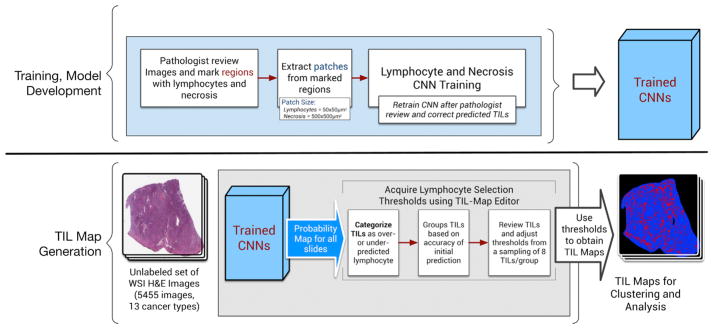
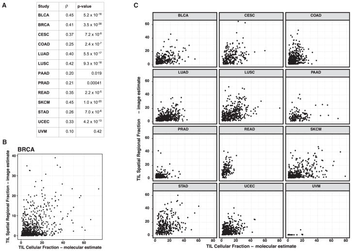
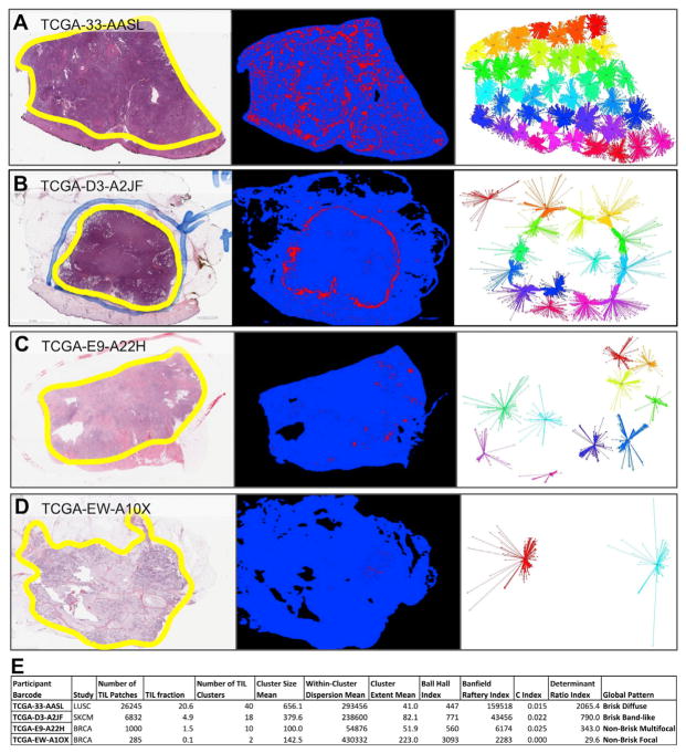
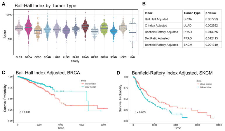
Beyond sample curation and basic pathologic characterization, the digitized H&E-stained images of TCGA samples remain underutilized. To highlight this resource, we present mappings of tumor-infiltrating lymphocytes (TILs) based on H&E images from 13 TCGA tumor types. These TIL maps are derived through computational staining using a convolutional neural network trained to classify patches of images. Affinity propagation revealed local spatial structure in TIL patterns and correlation with overall survival. TIL map structural patterns were grouped using standard histopathological parameters. These patterns are enriched in particular T cell subpopulations derived from molecular measures. TIL densities and spatial structure were differentially enriched among tumor types, immune subtypes, and tumor molecular subtypes, implying that spatial infiltrate state could reflect particular tumor cell aberration states. Obtaining spatial lymphocytic patterns linked to the rich genomic characterization of TCGA samples demonstrates one use for the TCGA image archives with insights into the tumor-immune microenvironment.
除了样本策展和基本病理特征描述,TCGA 样本的数字化 H&E 染色图像仍未得到充分利用。为了突出这一资源,我们根据 13 种 TCGA 肿瘤类型的 H&E 图像展示了肿瘤浸润淋巴细胞(TILs)的图谱。这些 TIL 图谱是通过使用经过训练以对图像块进行分类的卷积神经网络进行计算染色得到的。亲和传播揭示了 TIL 模式中的局部空间结构以及与总生存期的相关性。使用标准组织病理学参数对 TIL 图谱的结构模式进行分组。这些模式富含源自分子测量的特定 T 细胞亚群。TIL 密度和空间结构在肿瘤类型、免疫亚型和肿瘤分子亚型之间存在差异,这表明空间浸润状态可能反映特定的肿瘤细胞异常状态。获得与 TCGA 样本丰富基因组特征相关的空间淋巴细胞模式,展示了 TCGA 图像档案的一种用途,深入了解肿瘤免疫微环境。