Bauer Christopher E, Zachariou Valentinos, Seago Elayna, Gold Brian T
Department of Neuroscience, University of Kentucky, Lexington, KY, United States.
Sanders-Brown Center on Aging, University of Kentucky, Lexington, KY, United States.
Front Aging Neurosci. 2021 Jul 5;13:617947. doi: 10.3389/fnagi.2021.617947. eCollection 2021.
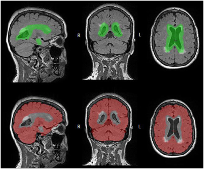
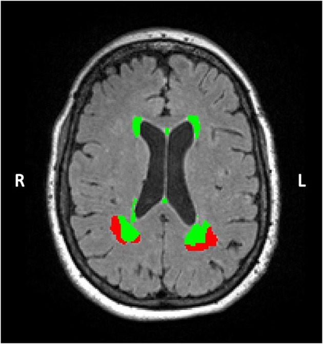
Cerebral white matter hyperintensities (WMHs) represent macrostructural brain damage associated with various etiologies. However, the relative contributions of various etiologies to WMH volume, as assessed via different neuroimaging measures, is not well-understood. Here, we explored associations between three potential early markers of white matter hyperintensity volume. Specifically, the unique variance in total and regional WMH volumes accounted for by white matter microstructure, brain iron concentration and cerebral blood flow (CBF) was assessed. Regional volumes explored were periventricular and deep regions. Eighty healthy older adults (ages 60-86) were scanned at 3 Tesla MRI using fluid-attenuated inversion recovery, diffusion tensor imaging (DTI), multi-echo gradient-recalled echo and pseudo-continuous arterial spin labeling sequences. In a stepwise regression model, DTI-based radial diffusivity accounted for significant variance in total WMH volume (adjusted change = 0.136). In contrast, iron concentration (adjusted change = 0.043) and CBF (adjusted change = 0.027) made more modest improvements to the variance accounted for in total WMH volume. However, there was an interaction between iron concentration and location on WMH volume such that iron concentration predicted deep ( = 0.034) but not periventricular ( = 0.414) WMH volume. Our results suggest that WM microstructure may be a better predictor of WMH volume than either brain iron or CBF but also draws attention to the possibility that some early WMH markers may be location-specific.
脑白质高信号(WMHs)代表与多种病因相关的大脑宏观结构损伤。然而,通过不同神经影像学测量评估的各种病因对WMH体积的相对贡献尚未得到充分理解。在此,我们探讨了白质高信号体积的三种潜在早期标志物之间的关联。具体而言,评估了白质微观结构、脑铁浓度和脑血流量(CBF)在总WMH体积和区域WMH体积中所占的独特方差。所探讨的区域体积为脑室周围和深部区域。使用液体衰减反转恢复序列、扩散张量成像(DTI)、多回波梯度回波序列和伪连续动脉自旋标记序列,对80名健康老年人(年龄60 - 86岁)进行3特斯拉磁共振成像扫描。在逐步回归模型中,基于DTI的径向扩散率在总WMH体积中占显著方差(调整后变化 = 0.136)。相比之下,铁浓度(调整后变化 = 0.043)和CBF(调整后变化 = 0.027)对总WMH体积中所占方差的改善较为有限。然而,铁浓度与WMH体积的位置之间存在相互作用,即铁浓度可预测深部WMH体积( = 0.034),但不能预测脑室周围WMH体积( = 0.414)。我们的结果表明,与脑铁或CBF相比,白质微观结构可能是WMH体积更好的预测指标,但也提请注意一些早期WMH标志物可能具有位置特异性的可能性。