Department of Surgical Sciences and Integrated Diagnostics, University of Genoa, 16126 Genoa, Italy.
Anesthesia and Intensive Care, San Martino Policlinico Hospital, IRCCS for Oncology and Neurosciences, 16132 Genoa, Italy.
Int J Mol Sci. 2021 Jul 13;22(14):7498. doi: 10.3390/ijms22147498.
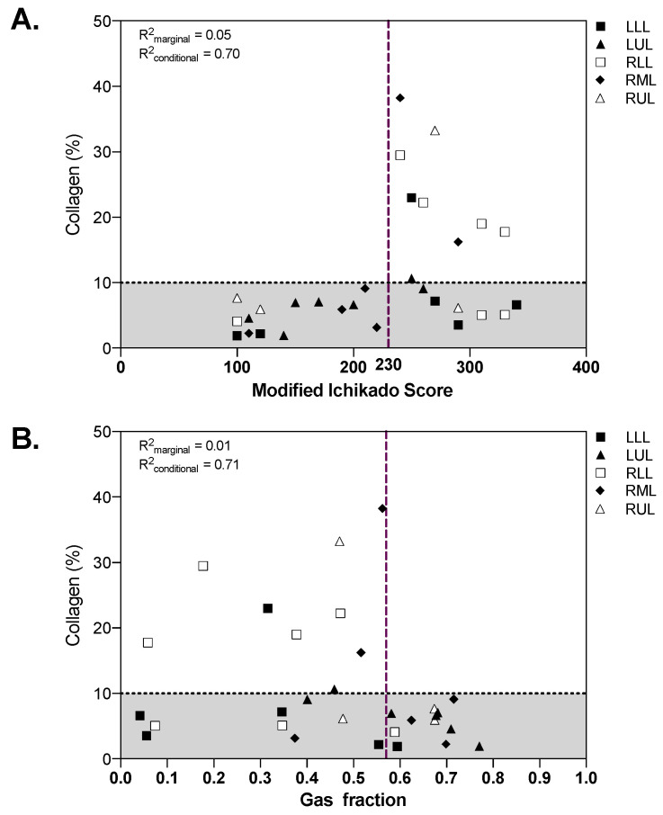
Lung fibrosis has specific computed tomography (CT) findings and represents a common finding in advanced COVID-19 pneumonia whose reversibility has been poorly investigated. The aim of this study was to quantify the extension of collagen deposition and aeration in postmortem cryobiopsies of critically ill COVID-19 patients and to describe the correlations with qualitative and quantitative analyses of lung CT. Postmortem transbronchial cryobiopsy samples were obtained, formalin fixed, paraffin embedded and stained with Sirius red to quantify collagen deposition, defining fibrotic samples as those with collagen deposition above 10%. Lung CT images were analyzed qualitatively with a radiographic score and quantitatively with computer-based analysis at the lobe level. Thirty samples from 10 patients with COVID-19 pneumonia deceased during invasive mechanical ventilation were included in this study. The median [interquartile range] percent collagen extension was 6.8% (4.6-16.2%). In fibrotic compared to nonfibrotic samples, the qualitative score was higher (260 (250-290) vs. 190 (120-270), = 0.036) while the gas fraction was lower (0.46 (0.32-0.47) vs. 0.59 (0.37-0.68), = 0.047). A radiographic score above 230 had 100% sensitivity (95% confidence interval, CI: 66.4% to 100%) and 66.7% specificity (95% CI: 41.0% to 92.3%) to detect fibrotic samples, while a gas fraction below 0.57 had 100% sensitivity (95% CI: 66.4% to 100%) and 57.1% specificity (95% CI: 26.3% to 88.0%). In COVID-19 pneumonia, qualitative and quantitative analyses of lung CT images have high sensitivity but moderate to low specificity to detect histopathological fibrosis. Pseudofibrotic CT findings do not always correspond to increased collagen deposition.
肺纤维化具有特定的计算机断层扫描(CT)表现,是 COVID-19 肺炎晚期的常见表现,但对其可逆性研究甚少。本研究旨在定量评估危重症 COVID-19 患者死后经支气管冷冻活检的胶原沉积和充气程度,并描述其与肺 CT 的定性和定量分析的相关性。对 10 例因接受有创机械通气而死亡的 COVID-19 肺炎患者的经支气管冷冻活检样本进行了研究,使用天狼星红染色进行胶原沉积定量,将胶原沉积超过 10%的样本定义为纤维化样本。对肺 CT 图像进行定性分析,采用放射学评分;对肺叶水平进行基于计算机的定量分析。本研究纳入了 30 例 COVID-19 肺炎患者的冷冻活检样本,这些患者在接受有创机械通气期间死亡。纤维化样本的胶原延伸中位数(四分位距)为 6.8%(4.6-16.2%)。与非纤维化样本相比,纤维化样本的定性评分更高(260(250-290)比 190(120-270), = 0.036),而充气分数更低(0.46(0.32-0.47)比 0.59(0.37-0.68), = 0.047)。放射学评分高于 230 时,检测纤维化样本的敏感性为 100%(95%置信区间:66.4%至 100%),特异性为 66.7%(95%置信区间:41.0%至 92.3%);充气分数低于 0.57 时,检测纤维化样本的敏感性为 100%(95%置信区间:66.4%至 100%),特异性为 57.1%(95%置信区间:26.3%至 88.0%)。在 COVID-19 肺炎中,肺 CT 图像的定性和定量分析具有较高的敏感性,但特异性为中等至较低,可用于检测组织病理学纤维化。假性纤维化 CT 表现并不总是与胶原沉积增加相对应。