Department of Liver Surgery, Liver Transplantation Center, West China Hospital of Sichuan University, No. 37 GuoXue Alley, Chengdu, 610041, People's Republic of China.
West China School of Medicine, West China Hospital, Sichuan University, No. 37 GuoXue Alley, Chengdu, 610041, People's Republic of China.
World J Surg Oncol. 2021 Aug 1;19(1):45. doi: 10.1186/s12957-021-02162-0.
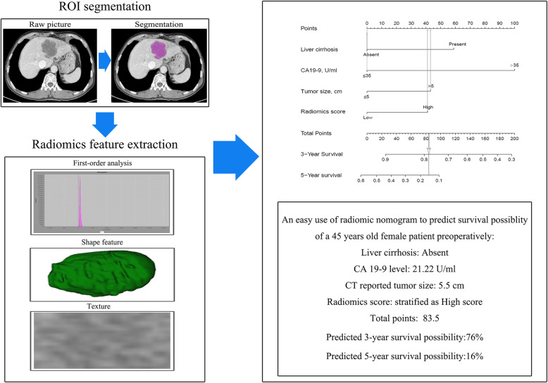
Intrahepatic cholangiocarcinoma is an aggressive liver carcinoma with increasing incidence and mortality. A good auxiliary prognostic prediction tool is desperately needed for the development of treatment strategies. The purpose of this study was to explore the prognostic value of the radiomics nomogram based on enhanced CT in intrahepatic cholangiocarcinoma.
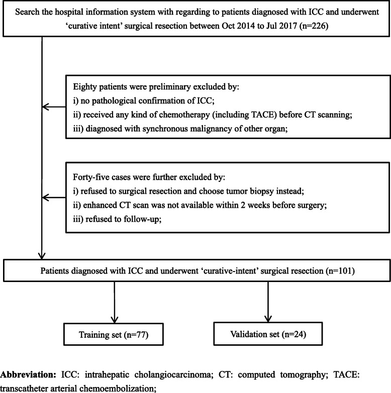
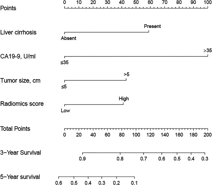
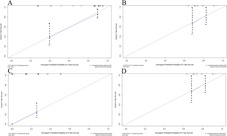
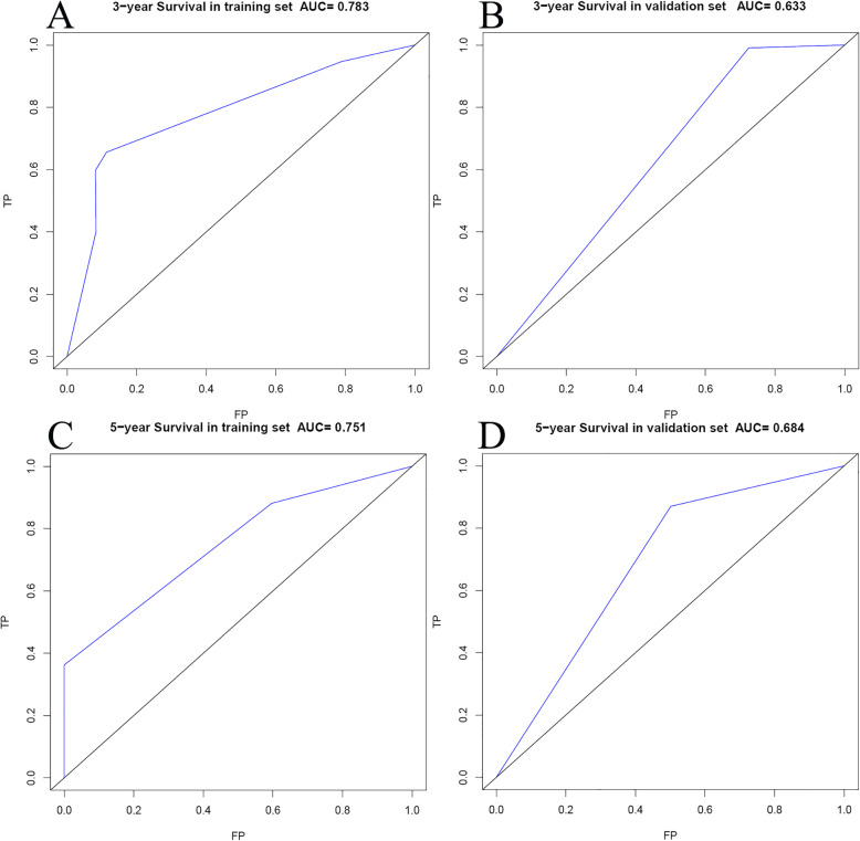
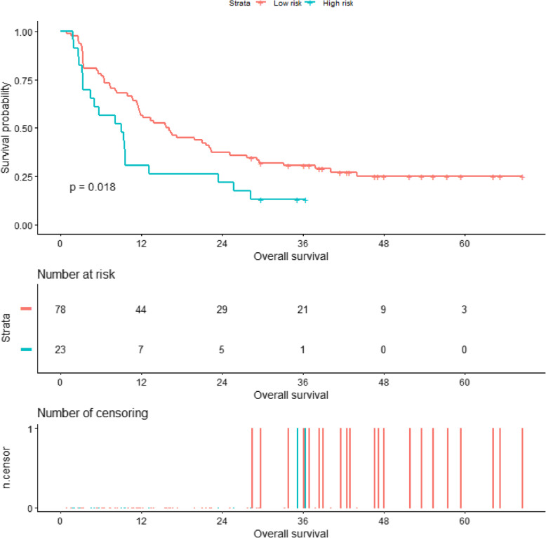
In this retrospective study, 101 patients with pathological confirmation of intrahepatic cholangiocarcinoma were recruited. A radiomics nomogram was developed by radiomics score and independent clinical risk factors selecting from multivariate Cox regression. All patients were stratified as high risk and low risk by a nomogram. Model performance and clinical usefulness were assessed by calibration curve, ROC curve, and survival curve.
A total of 101patients (mean age, 58.2 years old; range 36-79 years old) were included in the study. The 1-year, 3-year, and 5-year overall survival rates were 49.5%, 26.6%, and 14.4%, respectively, with a median survival time of 12.2 months in the whole set. The least absolute shrinkage and selection operator (LASSO) method selected 3 features. Multivariate Cox analysis found three independent prognostic factors. The radiomics nomogram showed a significant prognosis value with overall survival. There was a significant difference in the 1-year and 3-year survival rates of stratified high-risk and low-risk patients in the whole set (30.4% vs. 56.4% and 13.0% vs. 30.6%, respectively, p = 0.018).
This radiomics nomogram has potential application value in the preoperative prognostic prediction of intrahepatic cholangiocarcinoma and may facilitate in clinical decision-making.
肝内胆管细胞癌是一种侵袭性肝癌,其发病率和死亡率呈上升趋势。迫切需要一种良好的辅助预后预测工具来制定治疗策略。本研究旨在探讨基于增强 CT 的影像组学列线图在肝内胆管细胞癌中的预后价值。
在这项回顾性研究中,共纳入了 101 例经病理证实的肝内胆管细胞癌患者。通过多元 Cox 回归筛选影像组学评分和独立临床危险因素,建立影像组学列线图。根据列线图将所有患者分为高危组和低危组。通过校准曲线、ROC 曲线和生存曲线评估模型性能和临床实用性。
共纳入 101 例患者(平均年龄 58.2 岁,范围 36-79 岁)。整体 1 年、3 年和 5 年生存率分别为 49.5%、26.6%和 14.4%,中位总生存时间为 12.2 个月。最小绝对收缩和选择算子(LASSO)方法选择了 3 个特征。多因素 Cox 分析发现了 3 个独立的预后因素。影像组学列线图在总生存期方面具有显著的预后价值。在整个队列中,高危和低危分层患者的 1 年和 3 年生存率存在显著差异(分别为 30.4%比 56.4%和 13.0%比 30.6%,p=0.018)。
该影像组学列线图具有预测肝内胆管细胞癌术前预后的潜在应用价值,可能有助于临床决策。