Silverstein Steven M, Lai Adriann, Green Kyle M, Crosta Christen, Fradkin Samantha I, Ramchandran Rajeev S
Department of Psychiatry, University of Rochester Medical Center, Rochester, NY, USA.
Department of Neuroscience, University of Rochester Medical Center, Rochester, NY, USA.
Eye Brain. 2021 Jul 24;13:205-217. doi: 10.2147/EB.S317186. eCollection 2021.
Schizophrenia is associated with alterations in neural structure and function of the retina that are similar to changes seen in the retina and brain in multiple neurodegenerative disorders. Preliminary evidence suggests that retinal microvasculature may also be compromised in schizophrenia. The goal of this study was to determine, using optical coherence tomography angiography (OCTA), whether 1) schizophrenia is associated with alterations in retinal microvasculature density; and 2) microvasculature reductions are associated with retinal neural layer thinning and performance on a measure of verbal IQ.
Twenty-eight outpatients with schizophrenia or schizoaffective disorder and 37 psychiatrically healthy control subjects completed OCT and OCTA exams, and the Wechsler Test of Adult Reading.
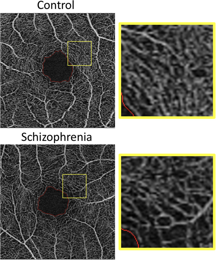
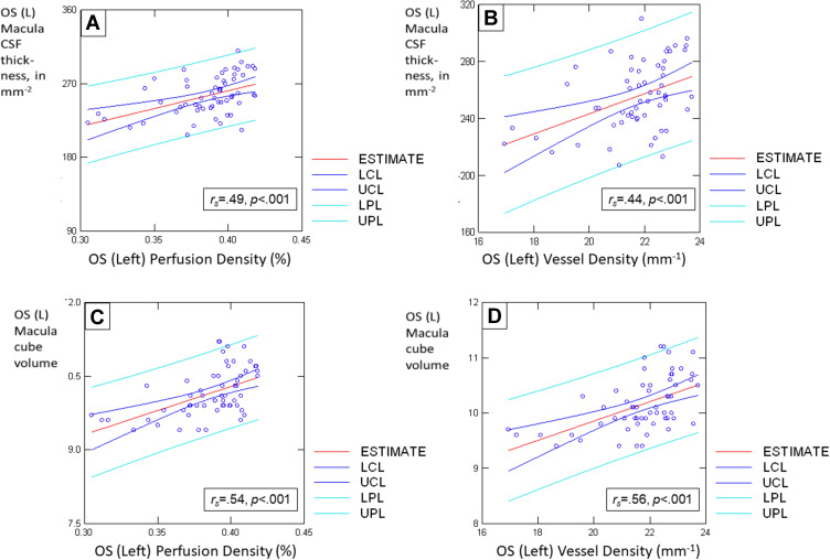
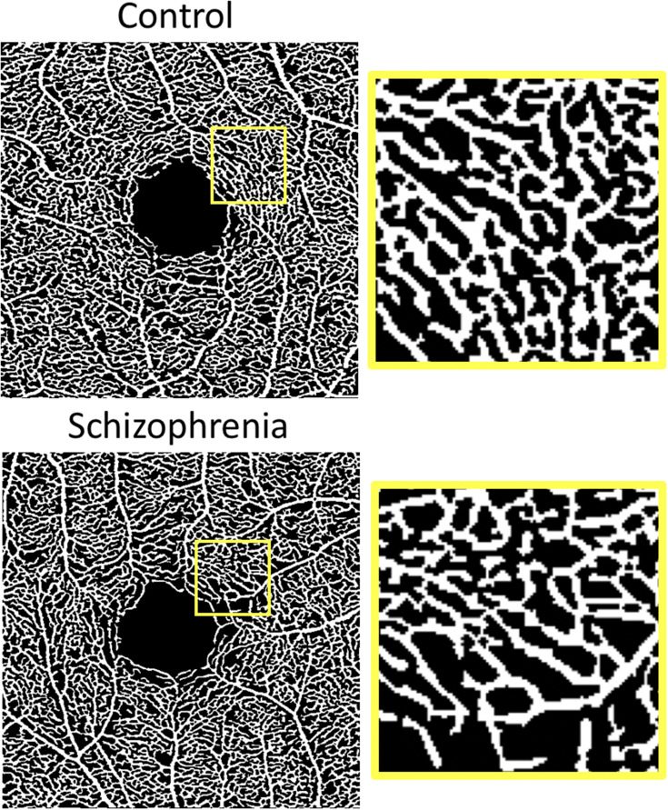
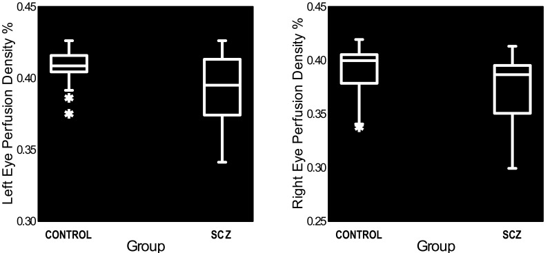
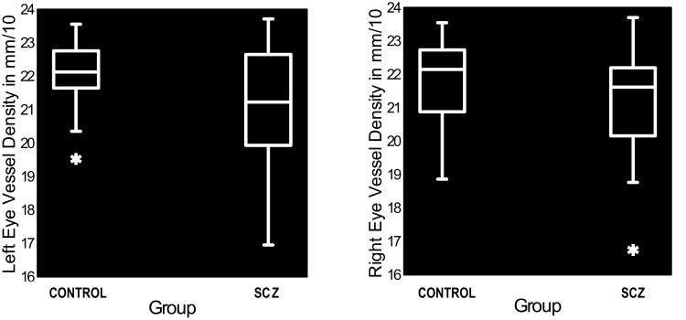
Schizophrenia patients were characterized by retinal microvasculature density reductions, and enlarged foveal avascular zones, in both eyes. These microvascular abnormalities were generally associated with thinning of retinal neural (macular and peripapillary nerve fiber layer) tissue (but the data were stronger for the left than the right eye) and lower scores on a proxy measure of verbal IQ. First- and later-episode patients did not differ significantly on OCTA findings.
The retinal microvasculature impairments seen in schizophrenia appear to be a biomarker of overall brain health, as is the case for multiple neurological conditions. Additional research is needed, however, to clarify contributions of social disadvantage and medical comorbidities to the findings.
精神分裂症与视网膜神经结构和功能的改变有关,这些改变类似于多种神经退行性疾病中视网膜和大脑所出现的变化。初步证据表明,精神分裂症患者的视网膜微血管系统也可能受到损害。本研究的目的是使用光学相干断层扫描血管造影(OCTA)来确定:1)精神分裂症是否与视网膜微血管密度改变有关;2)微血管减少是否与视网膜神经层变薄以及言语智商测量表现有关。
28例精神分裂症或分裂情感性障碍门诊患者以及37名精神健康的对照受试者完成了OCT和OCTA检查,以及韦氏成人阅读测验。
精神分裂症患者的双眼均表现为视网膜微血管密度降低以及中心凹无血管区扩大。这些微血管异常通常与视网膜神经(黄斑和视乳头周围神经纤维层)组织变薄有关(但左眼的数据比右眼更强),并且在言语智商的替代测量中得分较低。首发和复发患者在OCTA检查结果上没有显著差异。
精神分裂症患者出现的视网膜微血管损伤似乎是整体大脑健康的生物标志物,这与多种神经系统疾病的情况相同。然而,需要进一步的研究来阐明社会劣势和合并症对这些结果的影响。