Ammari Samy, Sallé de Chou Raoul, Assi Tarek, Touat Mehdi, Chouzenoux Emilie, Quillent Arnaud, Limkin Elaine, Dercle Laurent, Hadchiti Joya, Elhaik Mickael, Moalla Salma, Khettab Mohamed, Balleyguier Corinne, Lassau Nathalie, Dumont Sarah, Smolenschi Cristina
Biomaps, UMR1281 INSERM, CEA, CNRS, Université Paris-Saclay, 94805 Villejuif, France.
Department of Imaging, Gustave Roussy, Université Paris Saclay, 94805 Villejuif, France.
Diagnostics (Basel). 2021 Jul 14;11(7):1263. doi: 10.3390/diagnostics11071263.
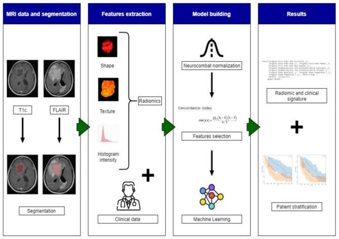
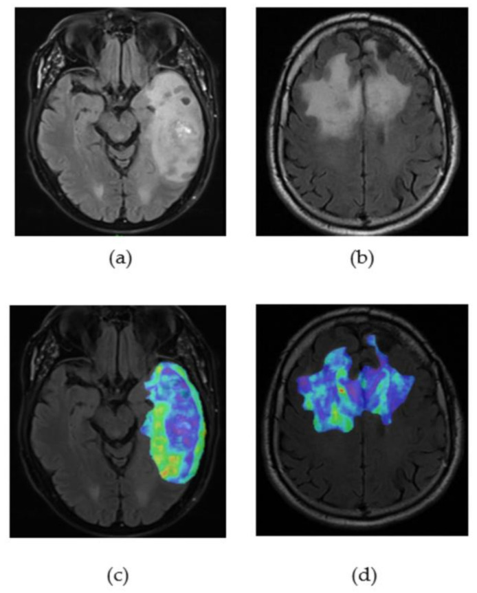
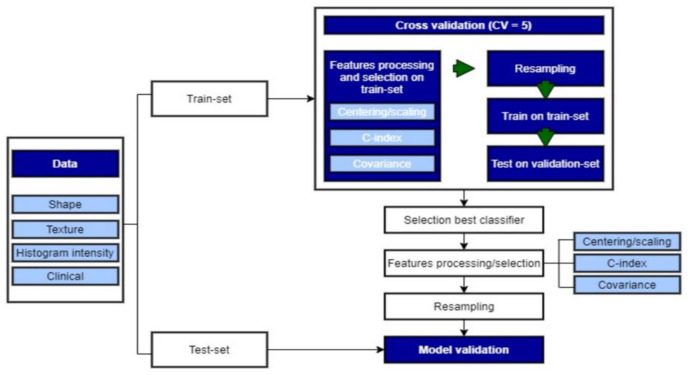
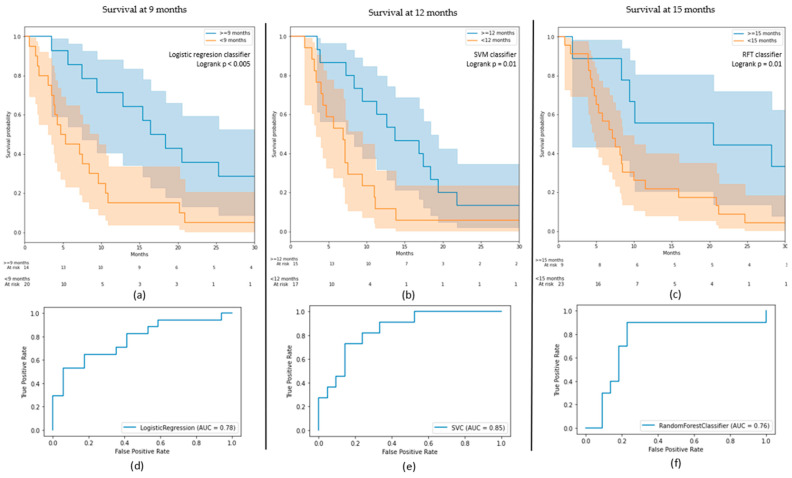
Anti-angiogenic therapy with bevacizumab is a widely used therapeutic option for recurrent glioblastoma (GBM). Nevertheless, the therapeutic response remains highly heterogeneous among GBM patients with discordant outcomes. Recent data have shown that radiomics, an advanced recent imaging analysis method, can help to predict both prognosis and therapy in a multitude of solid tumours. The objective of this study was to identify novel biomarkers, extracted from MRI and clinical data, which could predict overall survival (OS) and progression-free survival (PFS) in GBM patients treated with bevacizumab using machine-learning algorithms. In a cohort of 194 recurrent GBM patients (age range 18-80), radiomics data from pre-treatment T2 FLAIR and gadolinium-injected MRI images along with clinical features were analysed. Binary classification models for OS at 9, 12, and 15 months were evaluated. Our classification models successfully stratified the OS. The AUCs were equal to 0.78, 0.85, and 0.76 on the test sets (0.79, 0.82, and 0.87 on the training sets) for the 9-, 12-, and 15-month endpoints, respectively. Regressions yielded a C-index of 0.64 (0.74) for OS and 0.57 (0.69) for PFS. These results suggest that radiomics could assist in the elaboration of a predictive model for treatment selection in recurrent GBM patients.
贝伐单抗抗血管生成疗法是复发性胶质母细胞瘤(GBM)广泛应用的治疗选择。然而,GBM患者的治疗反应仍高度异质性,预后不一。近期数据表明,放射组学作为一种先进的影像分析方法,可帮助预测多种实体瘤的预后和治疗效果。本研究的目的是从MRI和临床数据中识别新的生物标志物,使用机器学习算法预测接受贝伐单抗治疗的GBM患者的总生存期(OS)和无进展生存期(PFS)。在一个包含194例复发性GBM患者(年龄范围18 - 80岁)的队列中,分析了治疗前T2 FLAIR和钆增强MRI图像的放射组学数据以及临床特征。评估了9个月、12个月和15个月时OS的二元分类模型。我们的分类模型成功地对OS进行了分层。在9个月、12个月和15个月终点的测试集上,AUC分别等于0.78、0.85和0.76(训练集上分别为0.79、0.82和0.87)。回归分析得出OS的C指数为0.64(0.74),PFS的C指数为0.57(0.69)。这些结果表明,放射组学有助于为复发性GBM患者制定治疗选择的预测模型。