• 文献检索
  • 文档翻译
  • 深度研究
  • 学术资讯
  • Suppr Zotero 插件Zotero 插件
  • 邀请有礼
  • 套餐&价格
  • 历史记录
应用&插件
Suppr Zotero 插件Zotero 插件浏览器插件Mac 客户端Windows 客户端微信小程序
定价
高级版会员购买积分包购买API积分包
服务
文献检索文档翻译深度研究API 文档MCP 服务
关于我们
关于 Suppr公司介绍联系我们用户协议隐私条款
关注我们

Suppr 超能文献

核心技术专利:CN118964589B侵权必究
粤ICP备2023148730 号-1Suppr @ 2026

文献检索

告别复杂PubMed语法,用中文像聊天一样搜索,搜遍4000万医学文献。AI智能推荐,让科研检索更轻松。

立即免费搜索

文件翻译

保留排版,准确专业,支持PDF/Word/PPT等文件格式,支持 12+语言互译。

免费翻译文档

深度研究

AI帮你快速写综述,25分钟生成高质量综述,智能提取关键信息,辅助科研写作。

立即免费体验

心肌细胞在感染 SARS-CoV-2 时通过分泌 CCL2 招募单核细胞。

Cardiomyocytes recruit monocytes upon SARS-CoV-2 infection by secreting CCL2.

机构信息

Department of Surgery, Weill Cornell Medicine, 1300 York Avenue, New York, NY 10065, USA.

Department of Microbiology, Icahn School of Medicine at Mount Sinai, 1468 Madison Avenue, New York, NY 10029, USA.

出版信息

Stem Cell Reports. 2021 Sep 14;16(9):2274-2288. doi: 10.1016/j.stemcr.2021.07.012. Epub 2021 Jul 20.

DOI:10.1016/j.stemcr.2021.07.012
PMID:34403650
原文链接:https://pmc.ncbi.nlm.nih.gov/articles/PMC8289700/
Abstract

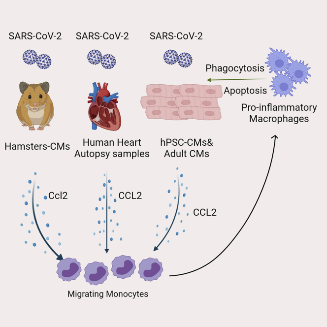
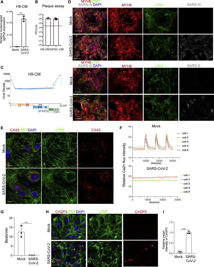
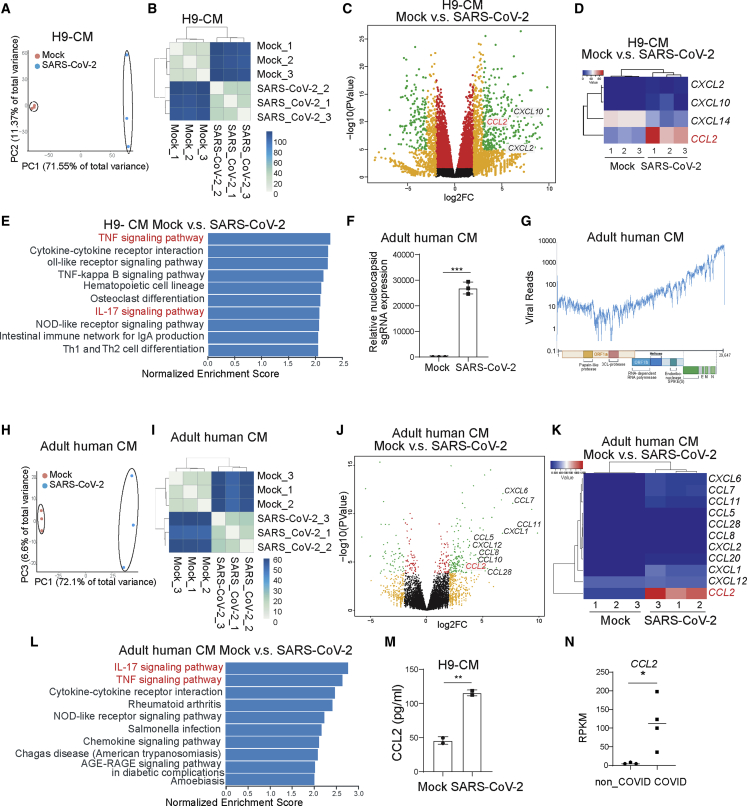
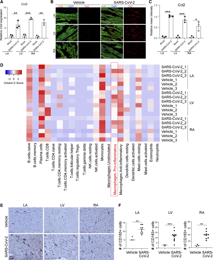
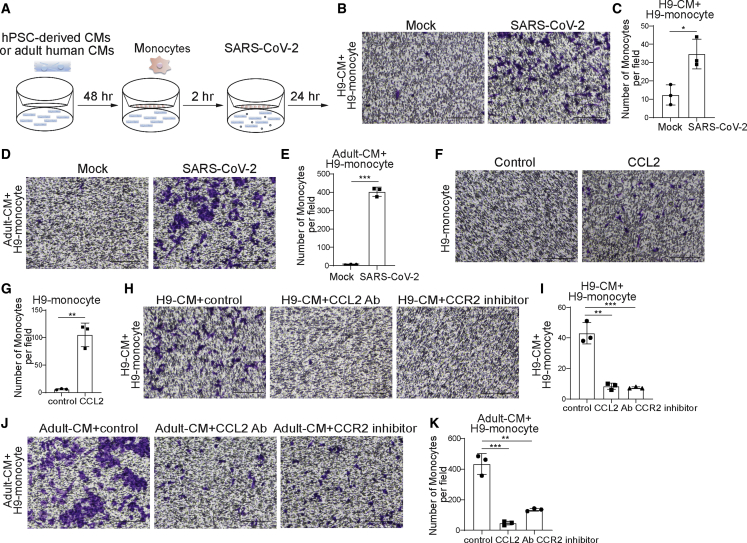
Heart injury has been reported in up to 20% of COVID-19 patients, yet the cause of myocardial histopathology remains unknown. Here, using an established in vivo hamster model, we demonstrate that SARS-CoV-2 can be detected in cardiomyocytes of infected animals. Furthermore, we found damaged cardiomyocytes in hamsters and COVID-19 autopsy samples. To explore the mechanism, we show that both human pluripotent stem cell-derived cardiomyocytes (hPSC-derived CMs) and adult cardiomyocytes (CMs) can be productively infected by SARS-CoV-2, leading to secretion of the monocyte chemoattractant cytokine CCL2 and subsequent monocyte recruitment. Increased CCL2 expression and monocyte infiltration was also observed in the hearts of infected hamsters. Although infected CMs suffer damage, we find that the presence of macrophages significantly reduces SARS-CoV-2-infected CMs. Overall, our study provides direct evidence that SARS-CoV-2 infects CMs in vivo and suggests a mechanism of immune cell infiltration and histopathology in heart tissues of COVID-19 patients.

摘要

高达 20%的 COVID-19 患者出现了心脏损伤,但心肌组织病理学的病因仍不清楚。在这里,我们使用已建立的体内仓鼠模型,证明了 SARS-CoV-2 可以在感染动物的心肌细胞中检测到。此外,我们在感染 SARS-CoV-2 的仓鼠和 COVID-19 尸检样本中发现了受损的心肌细胞。为了探讨其机制,我们发现人类多能干细胞来源的心肌细胞(hPSC 衍生的 CMs)和成年心肌细胞(CMs)均可被 SARS-CoV-2 有效感染,导致单核细胞趋化因子 CCL2 的分泌和随后的单核细胞募集。在感染的仓鼠心脏中也观察到 CCL2 表达增加和单核细胞浸润。尽管受感染的 CMs 受到损伤,但我们发现巨噬细胞的存在显著减少了 SARS-CoV-2 感染的 CMs。总的来说,我们的研究提供了直接证据,表明 SARS-CoV-2 在体内感染了 CMs,并提出了 COVID-19 患者心脏组织中免疫细胞浸润和组织病理学的一种机制。

https://cdn.ncbi.nlm.nih.gov/pmc/blobs/fcdb/8452518/46d5ecbe4a63/gr7.jpg
https://cdn.ncbi.nlm.nih.gov/pmc/blobs/fcdb/8452518/9ead71d88df6/fx1.jpg
https://cdn.ncbi.nlm.nih.gov/pmc/blobs/fcdb/8452518/1520a834153a/gr1.jpg
https://cdn.ncbi.nlm.nih.gov/pmc/blobs/fcdb/8452518/3f2460cf623e/gr2.jpg
https://cdn.ncbi.nlm.nih.gov/pmc/blobs/fcdb/8452518/f88c99ec2fb7/gr3.jpg
https://cdn.ncbi.nlm.nih.gov/pmc/blobs/fcdb/8452518/ad6fad78fb80/gr4.jpg
https://cdn.ncbi.nlm.nih.gov/pmc/blobs/fcdb/8452518/1aeaf8409cd2/gr5.jpg
https://cdn.ncbi.nlm.nih.gov/pmc/blobs/fcdb/8452518/eea767c0063c/gr6.jpg
https://cdn.ncbi.nlm.nih.gov/pmc/blobs/fcdb/8452518/46d5ecbe4a63/gr7.jpg

相似文献

1
Cardiomyocytes recruit monocytes upon SARS-CoV-2 infection by secreting CCL2.心肌细胞在感染 SARS-CoV-2 时通过分泌 CCL2 招募单核细胞。
Stem Cell Reports. 2021 Sep 14;16(9):2274-2288. doi: 10.1016/j.stemcr.2021.07.012. Epub 2021 Jul 20.
2
SARS-CoV-2 Infected Cardiomyocytes Recruit Monocytes by Secreting CCL2.严重急性呼吸综合征冠状病毒2感染的心肌细胞通过分泌CCL2招募单核细胞。
Res Sq. 2020 Nov 17:rs.3.rs-94634. doi: 10.21203/rs.3.rs-94634/v1.
3
Antiviral activity and safety of remdesivir against SARS-CoV-2 infection in human pluripotent stem cell-derived cardiomyocytes.瑞德西韦在人多能干细胞衍生的心肌细胞中抗 SARS-CoV-2 感染的抗病毒活性和安全性。
Antiviral Res. 2020 Dec;184:104955. doi: 10.1016/j.antiviral.2020.104955. Epub 2020 Oct 19.
4
Infiltrating monocytes drive cardiac dysfunction in a cardiomyocyte-restricted mouse model of SARS-CoV-2 infection.浸润单核细胞驱动 SARS-CoV-2 感染的心肌细胞特异性小鼠模型的心功能障碍。
J Virol. 2024 Sep 17;98(9):e0117924. doi: 10.1128/jvi.01179-24. Epub 2024 Aug 29.
5
Highly Efficient SARS-CoV-2 Infection of Human Cardiomyocytes: Spike Protein-Mediated Cell Fusion and Its Inhibition.高效感染人类心肌细胞的 SARS-CoV-2:刺突蛋白介导的细胞融合及其抑制。
J Virol. 2021 Nov 23;95(24):e0136821. doi: 10.1128/JVI.01368-21. Epub 2021 Oct 6.
6
Genome-wide analyses reveal the detrimental impacts of SARS-CoV-2 viral gene Orf9c on human pluripotent stem cell-derived cardiomyocytes.全基因组分析揭示了 SARS-CoV-2 病毒基因 Orf9c 对人多能干细胞衍生心肌细胞的有害影响。
Stem Cell Reports. 2022 Mar 8;17(3):522-537. doi: 10.1016/j.stemcr.2022.01.014. Epub 2022 Feb 17.
7
SARS-CoV-2 Infects Human Pluripotent Stem Cell-Derived Cardiomyocytes, Impairing Electrical and Mechanical Function.SARS-CoV-2 感染人多能干细胞衍生的心肌细胞,损害电和机械功能。
Stem Cell Reports. 2021 Mar 9;16(3):478-492. doi: 10.1016/j.stemcr.2021.02.008. Epub 2021 Feb 13.
8
Network pharmacology and RNA-sequencing reveal the molecular mechanism of Xuebijing injection on COVID-19-induced cardiac dysfunction.网络药理学和 RNA 测序揭示了血必净注射液治疗 COVID-19 相关性心功能障碍的分子机制。
Comput Biol Med. 2021 Apr;131:104293. doi: 10.1016/j.compbiomed.2021.104293. Epub 2021 Feb 22.
9
SARS-CoV-2 induces cardiomyocyte apoptosis and inflammation but can be ameliorated by ACE inhibitor Captopril.SARS-CoV-2 诱导心肌细胞凋亡和炎症,但可被 ACE 抑制剂卡托普利改善。
Antiviral Res. 2023 Jul;215:105636. doi: 10.1016/j.antiviral.2023.105636. Epub 2023 May 18.
10
SARS-CoV-2 infects and induces cytotoxic effects in human cardiomyocytes.SARS-CoV-2 感染并诱导人心肌细胞产生细胞毒性作用。
Cardiovasc Res. 2020 Dec 1;116(14):2207-2215. doi: 10.1093/cvr/cvaa267.

引用本文的文献

1
APOE genotype confers context-dependent neurovascular vulnerability in immune-vascularized human forebrain organoids.载脂蛋白E(APOE)基因型在免疫血管化的人类前脑类器官中赋予了依赖于背景的神经血管易损性。
bioRxiv. 2025 May 9:2025.05.08.652864. doi: 10.1101/2025.05.08.652864.
2
Immune modulation: the key to combat SARS-CoV-2 induced myocardial injury.免疫调节:对抗SARS-CoV-2诱导的心肌损伤的关键。
Front Immunol. 2025 May 14;16:1561946. doi: 10.3389/fimmu.2025.1561946. eCollection 2025.
3
Organoids in virology.病毒学中的类器官

本文引用的文献

1
SARS-CoV-2 Infects Human Pluripotent Stem Cell-Derived Cardiomyocytes, Impairing Electrical and Mechanical Function.SARS-CoV-2 感染人多能干细胞衍生的心肌细胞,损害电和机械功能。
Stem Cell Reports. 2021 Mar 9;16(3):478-492. doi: 10.1016/j.stemcr.2021.02.008. Epub 2021 Feb 13.
2
Evidence of SARS-CoV-2 Transcriptional Activity in Cardiomyocytes of COVID-19 Patients without Clinical Signs of Cardiac Involvement.新型冠状病毒肺炎(COVID-19)患者心肌细胞中存在严重急性呼吸综合征冠状病毒2(SARS-CoV-2)转录活性的证据,这些患者无心脏受累的临床体征。
Biomedicines. 2020 Dec 18;8(12):626. doi: 10.3390/biomedicines8120626.
3
Using Cardiovascular Cells from Human Pluripotent Stem Cells for COVID-19 Research: Why the Heart Fails.
Npj Viruses. 2024 Feb 7;2(1):5. doi: 10.1038/s44298-024-00017-5.
4
Animal Models of Non-Respiratory, Post-Acute Sequelae of COVID-19.新冠病毒病非呼吸道急性后遗症的动物模型
Viruses. 2025 Jan 14;17(1):98. doi: 10.3390/v17010098.
5
COVID-19: a multi-organ perspective.新型冠状病毒肺炎:多器官视角。
Front Cell Infect Microbiol. 2024 Oct 18;14:1425547. doi: 10.3389/fcimb.2024.1425547. eCollection 2024.
6
Chemokine CCL2 and its receptor CCR2 in different age groups of patients with COVID-19.新型冠状病毒肺炎患者不同年龄组趋化因子 CCL2 及其受体 CCR2 的表达。
BMC Immunol. 2024 Oct 26;25(1):72. doi: 10.1186/s12865-024-00662-8.
7
Possible mechanisms of SARS-CoV-2-associated myocardial fibrosis: reflections in the post-pandemic era.新型冠状病毒2型相关心肌纤维化的可能机制:大流行后时代的思考
Front Microbiol. 2024 Oct 8;15:1470953. doi: 10.3389/fmicb.2024.1470953. eCollection 2024.
8
The emerging role of heart-on-a-chip systems in delineating mechanisms of SARS-CoV-2-induced cardiac dysfunction.芯片心脏系统在阐明SARS-CoV-2诱导的心脏功能障碍机制中的新作用。
Bioeng Transl Med. 2023 Aug 8;9(3):e10581. doi: 10.1002/btm2.10581. eCollection 2024 May.
9
RNA m5C methylation modification: a potential therapeutic target for SARS-CoV-2-associated myocarditis.RNA m5C 甲基化修饰:SARS-CoV-2 相关心肌炎的潜在治疗靶点。
Front Immunol. 2024 Apr 23;15:1380697. doi: 10.3389/fimmu.2024.1380697. eCollection 2024.
10
The inflammatory spectrum of cardiomyopathies.心肌病的炎症谱
Front Cardiovasc Med. 2024 Feb 23;11:1251780. doi: 10.3389/fcvm.2024.1251780. eCollection 2024.
使用人类多能干细胞的心血管细胞进行 COVID-19 研究:心脏衰竭的原因。
Stem Cell Reports. 2021 Mar 9;16(3):385-397. doi: 10.1016/j.stemcr.2020.11.003. Epub 2020 Dec 10.
4
Identification of SARS-CoV-2 inhibitors using lung and colonic organoids.使用肺和结肠类器官鉴定 SARS-CoV-2 抑制剂。
Nature. 2021 Jan;589(7841):270-275. doi: 10.1038/s41586-020-2901-9. Epub 2020 Oct 28.
5
SARS-CoV-2 infects and induces cytotoxic effects in human cardiomyocytes.SARS-CoV-2 感染并诱导人心肌细胞产生细胞毒性作用。
Cardiovasc Res. 2020 Dec 1;116(14):2207-2215. doi: 10.1093/cvr/cvaa267.
6
Human iPSC-Derived Cardiomyocytes Are Susceptible to SARS-CoV-2 Infection.人诱导多能干细胞衍生的心肌细胞易受新冠病毒感染。
Cell Rep Med. 2020 Jul 21;1(4):100052. doi: 10.1016/j.xcrm.2020.100052. Epub 2020 Jun 29.
7
SARS-CoV-2 in cardiac tissue of a child with COVID-19-related multisystem inflammatory syndrome.新型冠状病毒肺炎相关多系统炎症综合征患儿心脏组织中的严重急性呼吸综合征冠状病毒2
Lancet Child Adolesc Health. 2020 Oct;4(10):790-794. doi: 10.1016/S2352-4642(20)30257-1. Epub 2020 Aug 20.
8
Association of Cardiac Infection With SARS-CoV-2 in Confirmed COVID-19 Autopsy Cases.心脏感染与确诊 COVID-19 尸检病例中 SARS-CoV-2 的关联。
JAMA Cardiol. 2020 Nov 1;5(11):1281-1285. doi: 10.1001/jamacardio.2020.3551.
9
Myocarditis in a 16-year-old boy positive for SARS-CoV-2.一名16岁SARS-CoV-2检测呈阳性男孩的心肌炎
Lancet. 2020 Jun 27;395(10242):e116. doi: 10.1016/S0140-6736(20)31307-6.
10
A Human Pluripotent Stem Cell-based Platform to Study SARS-CoV-2 Tropism and Model Virus Infection in Human Cells and Organoids.基于人多能干细胞的平台,用于研究 SARS-CoV-2 嗜性和人类细胞及类器官中的病毒感染模型。
Cell Stem Cell. 2020 Jul 2;27(1):125-136.e7. doi: 10.1016/j.stem.2020.06.015. Epub 2020 Jun 19.