Smith & Nephew Plc., Fort Worth, TX 76107, USA.
Int J Mol Sci. 2021 Aug 11;22(16):8643. doi: 10.3390/ijms22168643.
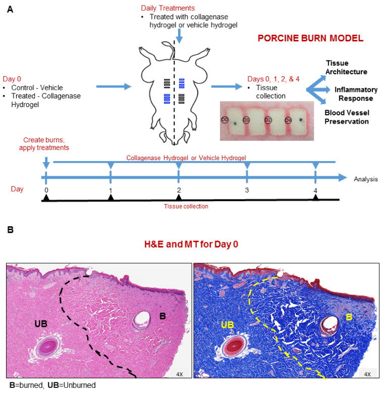
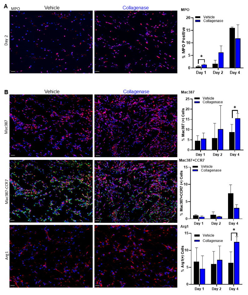
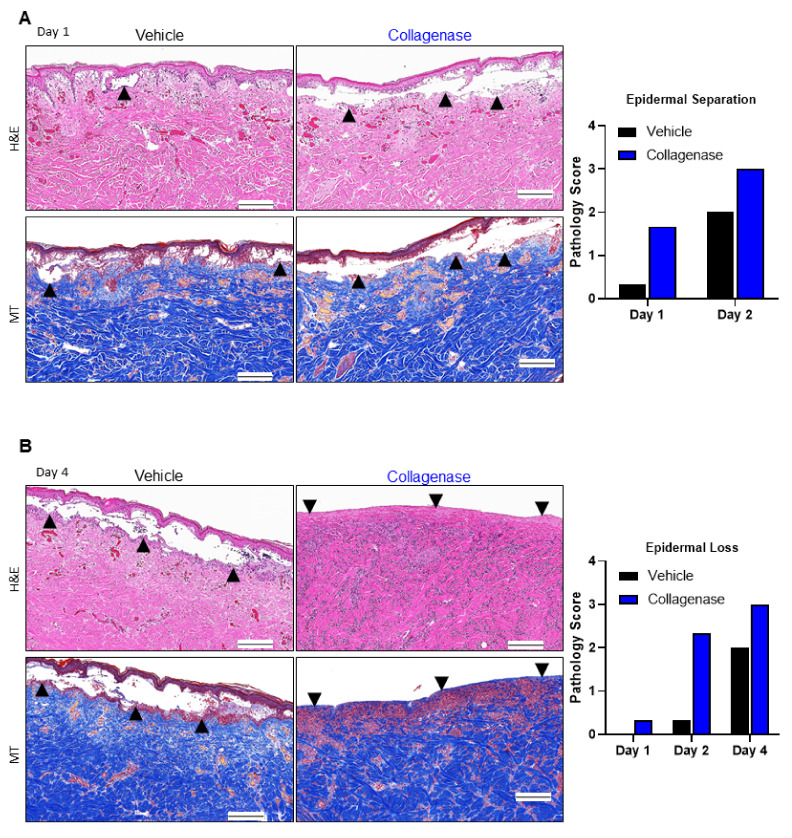
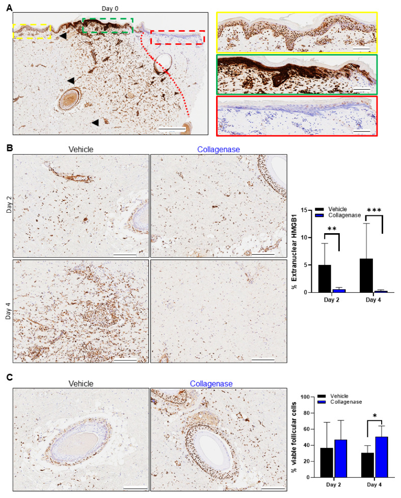
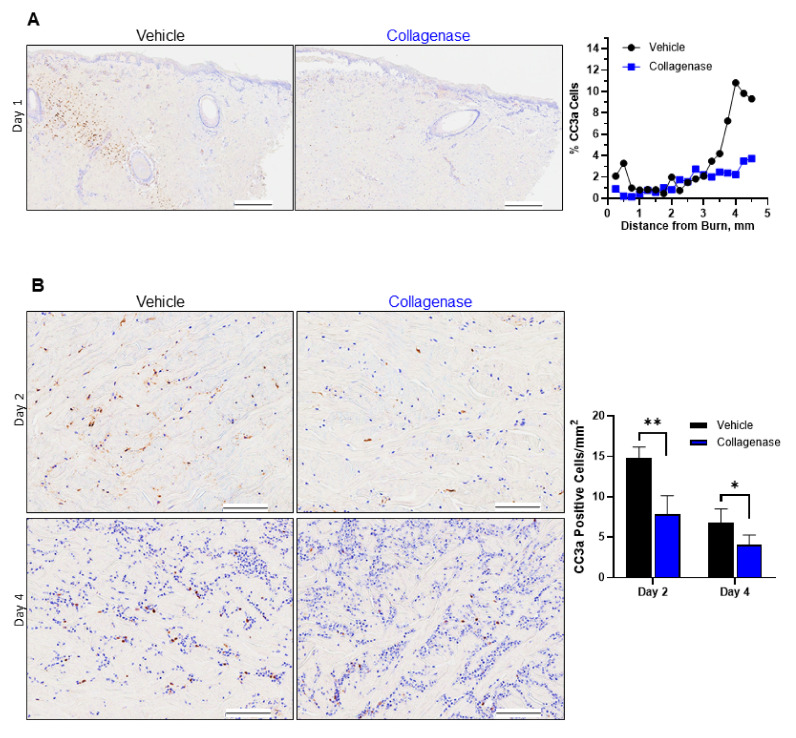
Clostridium collagenase has provided superior clinical results in achieving digestion of immediate and accumulating devitalized collagen tissue. Recent studies suggest that debridement via Clostridium collagenase modulates a cellular response to foster an anti-inflammatory microenvironment milieu, allowing for a more coordinated healing response. In an effort to better understand its role in burn wounds, we evaluated Clostridium collagenase's ability to effectively minimize burn progression using the classic burn comb model in pigs. Following burn injury, wounds were treated with Clostridium collagenase or control vehicle daily and biopsied at various time points. Biopsies were evaluated for factors associated with progressing necrosis as well as inflammatory response associated with treatment. Data presented herein showed that Clostridium collagenase treatment prevented destruction of dermal collagen. Additionally, treatment with collagenase reduced necrosis (HMGB1) and apoptosis (CC3a) early in burn injuries, allowing for increased infiltration of cells and protecting tissue from conversion. Furthermore, early epidermal separation and epidermal loss with a clearly defined basement membrane was observed in the treated wounds. We also show that collagenase treatment provided an early and improved inflammatory response followed by faster resolution in neutrophils. In assessing the inflammatory response, collagenase-treated wounds exhibited significantly greater neutrophil influx at day 1, with macrophage recruitment throughout days 2 and 4. In further evaluation, macrophage polarization to MHC II and vascular network maintenance were significantly increased in collagenase-treated wounds, indicative of a pro-resolving macrophage environment. Taken together, these data validate the impact of clostridial collagenases in the pathophysiology of burn wounds and that they complement patient outcomes in the clinical scenario.
胶原梭菌在实现即时消化和积累失活胶原组织方面提供了卓越的临床效果。最近的研究表明,胶原梭菌清创通过调节细胞反应来促进抗炎微环境,从而实现更协调的愈合反应。为了更好地了解其在烧伤创面中的作用,我们使用经典的烧伤复合模型在猪身上评估了胶原梭菌有效减轻烧伤进展的能力。烧伤后,每天用胶原梭菌或对照剂处理伤口,并在不同时间点进行活检。对活检进行评估,以确定与进展性坏死以及与治疗相关的炎症反应相关的因素。本文提供的数据表明,胶原梭菌处理可防止真皮胶原的破坏。此外,胶原酶治疗可减少烧伤早期的坏死(HMGB1)和凋亡(CC3a),从而增加细胞浸润,并防止组织转化。此外,在处理的伤口中观察到早期表皮分离和表皮丢失,并有明确的基底膜。我们还表明,胶原酶治疗可提供早期和改善的炎症反应,随后中性粒细胞更快消退。在评估炎症反应时,胶原酶处理的伤口在第 1 天表现出明显更大的中性粒细胞浸润,在第 2 天和第 4 天巨噬细胞募集。进一步评估表明,胶原酶处理的伤口中 MHC II 表达的巨噬细胞极化和血管网络维持显著增加,表明存在促解决的巨噬细胞环境。综上所述,这些数据验证了梭菌胶原酶在烧伤创面病理生理学中的作用,并且它们在临床情况下补充了患者的治疗效果。