• 文献检索
  • 文档翻译
  • 深度研究
  • 学术资讯
  • Suppr Zotero 插件Zotero 插件
  • 邀请有礼
  • 套餐&价格
  • 历史记录
应用&插件
Suppr Zotero 插件Zotero 插件浏览器插件Mac 客户端Windows 客户端微信小程序
定价
高级版会员购买积分包购买API积分包
服务
文献检索文档翻译深度研究API 文档MCP 服务
关于我们
关于 Suppr公司介绍联系我们用户协议隐私条款
关注我们

Suppr 超能文献

核心技术专利:CN118964589B侵权必究
粤ICP备2023148730 号-1Suppr @ 2026

文献检索

告别复杂PubMed语法,用中文像聊天一样搜索,搜遍4000万医学文献。AI智能推荐,让科研检索更轻松。

立即免费搜索

文件翻译

保留排版,准确专业,支持PDF/Word/PPT等文件格式,支持 12+语言互译。

免费翻译文档

深度研究

AI帮你快速写综述,25分钟生成高质量综述,智能提取关键信息,辅助科研写作。

立即免费体验

基于迁移学习的病理切片图像分类方法

Transfer Learning Approach for Classification of Histopathology Whole Slide Images.

机构信息

Department of Computer Science and Information Technology, University of Balochistan, Quetta 87300, Pakistan.

College of Computer Science and Information Systems, Najran University, Najran 61441, Saudi Arabia.

出版信息

Sensors (Basel). 2021 Aug 9;21(16):5361. doi: 10.3390/s21165361.

DOI:10.3390/s21165361
PMID:34450802
原文链接:https://pmc.ncbi.nlm.nih.gov/articles/PMC8401188/
Abstract

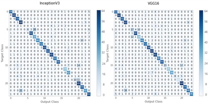
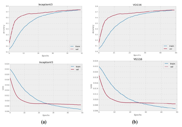
The classification of whole slide images (WSIs) provides physicians with an accurate analysis of diseases and also helps them to treat patients effectively. The classification can be linked to further detailed analysis and diagnosis. Deep learning (DL) has made significant advances in the medical industry, including the use of magnetic resonance imaging (MRI) scans, computerized tomography (CT) scans, and electrocardiograms (ECGs) to detect life-threatening diseases, including heart disease, cancer, and brain tumors. However, more advancement in the field of pathology is needed, but the main hurdle causing the slow progress is the shortage of large-labeled datasets of histopathology images to train the models. The Kimia Path24 dataset was particularly created for the classification and retrieval of histopathology images. It contains 23,916 histopathology patches with 24 tissue texture classes. A transfer learning-based framework is proposed and evaluated on two famous DL models, Inception-V3 and VGG-16. To improve the productivity of Inception-V3 and VGG-16, we used their pre-trained weights and concatenated these with an image vector, which is used as input for the training of the same architecture. Experiments show that the proposed innovation improves the accuracy of both famous models. The patch-to-scan accuracy of VGG-16 is improved from 0.65 to 0.77, and for the Inception-V3, it is improved from 0.74 to 0.79.

摘要

全切片图像 (WSI) 的分类为医生提供了对疾病的准确分析,也有助于他们有效地治疗患者。分类可以与进一步的详细分析和诊断相关联。深度学习 (DL) 在医疗行业取得了重大进展,包括使用磁共振成像 (MRI) 扫描、计算机断层扫描 (CT) 扫描和心电图 (ECG) 来检测危及生命的疾病,包括心脏病、癌症和脑肿瘤。然而,病理学领域需要更多的进展,但导致进展缓慢的主要障碍是缺乏用于训练模型的大型标记组织病理学图像数据集。Kimia Path24 数据集是专门为组织病理学图像的分类和检索而创建的。它包含 23916 个具有 24 种组织纹理类别的组织病理学斑块。提出并评估了基于迁移学习的框架,该框架在两个著名的深度学习模型 Inception-V3 和 VGG-16 上进行了评估。为了提高 Inception-V3 和 VGG-16 的生产力,我们使用了它们的预训练权重,并将其与图像向量串联,将其用作相同架构的训练输入。实验表明,所提出的创新提高了这两个著名模型的准确性。VGG-16 的斑块到扫描的准确率从 0.65 提高到 0.77,而对于 Inception-V3,则从 0.74 提高到 0.79。

https://cdn.ncbi.nlm.nih.gov/pmc/blobs/d1bb/8401188/cdb2bbb57d4d/sensors-21-05361-g004.jpg
https://cdn.ncbi.nlm.nih.gov/pmc/blobs/d1bb/8401188/41e0882ee3fa/sensors-21-05361-g001.jpg
https://cdn.ncbi.nlm.nih.gov/pmc/blobs/d1bb/8401188/20ce30a8cb7a/sensors-21-05361-g002.jpg
https://cdn.ncbi.nlm.nih.gov/pmc/blobs/d1bb/8401188/d4e2526444ff/sensors-21-05361-g003.jpg
https://cdn.ncbi.nlm.nih.gov/pmc/blobs/d1bb/8401188/cdb2bbb57d4d/sensors-21-05361-g004.jpg

相似文献

1
Transfer Learning Approach for Classification of Histopathology Whole Slide Images.基于迁移学习的病理切片图像分类方法
Sensors (Basel). 2021 Aug 9;21(16):5361. doi: 10.3390/s21165361.
2
Brain tumor classification in MRI image using convolutional neural network.基于卷积神经网络的MRI图像脑肿瘤分类
Math Biosci Eng. 2020 Sep 15;17(5):6203-6216. doi: 10.3934/mbe.2020328.
3
Automated classification of histopathology images using transfer learning.使用迁移学习对组织病理学图像进行自动分类。
Artif Intell Med. 2019 Nov;101:101743. doi: 10.1016/j.artmed.2019.101743. Epub 2019 Nov 3.
4
Deep Transfer Learning Approaches in Performance Analysis of Brain Tumor Classification Using MRI Images.基于 MRI 图像的脑肿瘤分类性能分析中的深度迁移学习方法。
J Healthc Eng. 2022 Mar 8;2022:3264367. doi: 10.1155/2022/3264367. eCollection 2022.
5
Pre-trained deep learning models for brain MRI image classification.用于脑磁共振成像(MRI)图像分类的预训练深度学习模型。
Front Hum Neurosci. 2023 Apr 20;17:1150120. doi: 10.3389/fnhum.2023.1150120. eCollection 2023.
6
Deep feature classification of angiomyolipoma without visible fat and renal cell carcinoma in abdominal contrast-enhanced CT images with texture image patches and hand-crafted feature concatenation.利用纹理图像补丁和手工特征串联对腹部增强 CT 图像中无可见脂肪的血管平滑肌脂肪瘤和肾细胞癌进行深度特征分类。
Med Phys. 2018 Apr;45(4):1550-1561. doi: 10.1002/mp.12828. Epub 2018 Mar 25.
7
MediNet: transfer learning approach with MediNet medical visual database.MediNet:基于MediNet医学视觉数据库的迁移学习方法。
Multimed Tools Appl. 2023 Mar 20:1-44. doi: 10.1007/s11042-023-14831-1.
8
Conventional Machine Learning versus Deep Learning for Magnification Dependent Histopathological Breast Cancer Image Classification: A Comparative Study with Visual Explanation.用于基于放大倍数的组织病理学乳腺癌图像分类的传统机器学习与深度学习:一项带有可视化解释的比较研究
Diagnostics (Basel). 2021 Mar 16;11(3):528. doi: 10.3390/diagnostics11030528.
9
Brain tumor classification for MR images using transfer learning and fine-tuning.基于迁移学习和微调的磁共振图像脑肿瘤分类。
Comput Med Imaging Graph. 2019 Jul;75:34-46. doi: 10.1016/j.compmedimag.2019.05.001. Epub 2019 May 18.
10
MRI-Based Brain Tumor Classification Using Ensemble of Deep Features and Machine Learning Classifiers.基于 MRI 的脑肿瘤分类:深度学习特征集与机器学习分类器的应用
Sensors (Basel). 2021 Mar 22;21(6):2222. doi: 10.3390/s21062222.

引用本文的文献

1
Colorectal cancer tumor grade segmentation: A new dataset and baseline results.结直肠癌肿瘤分级分割:一个新的数据集及基线结果。
Heliyon. 2025 Feb 11;11(4):e42467. doi: 10.1016/j.heliyon.2025.e42467. eCollection 2025 Feb 28.
2
Applicability of the regression approach for histological multi-class grading in clear cell renal cell carcinoma.回归方法在透明细胞肾细胞癌组织学多分类分级中的适用性。
Regen Ther. 2025 Jan 25;28:431-437. doi: 10.1016/j.reth.2025.01.011. eCollection 2025 Mar.
3
Digital pathology, deep learning, and cancer: a narrative review.

本文引用的文献

1
Brain tumor classification in MRI image using convolutional neural network.基于卷积神经网络的MRI图像脑肿瘤分类
Math Biosci Eng. 2020 Sep 15;17(5):6203-6216. doi: 10.3934/mbe.2020328.
2
Enhanced Image-Based Endoscopic Pathological Site Classification Using an Ensemble of Deep Learning Models.基于增强图像的内镜病理部位分类的深度学习模型集成方法。
Sensors (Basel). 2020 Oct 22;20(21):5982. doi: 10.3390/s20215982.
3
Breast Cancer Histopathology Image Classification Using an Ensemble of Deep Learning Models.基于深度学习模型集成的乳腺癌病理图像分类。
数字病理学、深度学习与癌症:一篇综述
Transl Cancer Res. 2024 May 31;13(5):2544-2560. doi: 10.21037/tcr-23-964. Epub 2024 May 22.
4
AI-Assisted Fusion of Scanning Electrochemical Microscopy Images Using Novel Soft Probe.使用新型软探针的扫描电化学显微镜图像的人工智能辅助融合
ACS Meas Sci Au. 2022 Aug 19;2(6):576-583. doi: 10.1021/acsmeasuresciau.2c00032. eCollection 2022 Dec 21.
5
Deep Domain Adversarial Learning for Species-Agnostic Classification of Histologic Subtypes of Osteosarcoma.基于深度领域对抗学习的骨肉瘤组织学亚型的物种不可知分类。
Am J Pathol. 2023 Jan;193(1):60-72. doi: 10.1016/j.ajpath.2022.09.009. Epub 2022 Oct 27.
6
Improving feature extraction from histopathological images through a fine-tuning ImageNet model.通过微调ImageNet模型改进从组织病理学图像中提取特征。
J Pathol Inform. 2022 Jun 30;13:100115. doi: 10.1016/j.jpi.2022.100115. eCollection 2022.
7
Perception without preconception: comparison between the human and machine learner in recognition of tissues from histological sections.无预设认知的感知:在识别组织切片方面,人与机器学习者的比较。
Sci Rep. 2022 Sep 30;12(1):16420. doi: 10.1038/s41598-022-20012-1.
8
Towards Interpretable Deep Learning: A Feature Selection Framework for Prognostics and Health Management Using Deep Neural Networks.迈向可解释的深度学习:基于深度神经网络的预测与健康管理的特征选择框架。
Sensors (Basel). 2021 Sep 1;21(17):5888. doi: 10.3390/s21175888.
Sensors (Basel). 2020 Aug 5;20(16):4373. doi: 10.3390/s20164373.
4
Deep Learning for Whole Slide Image Analysis: An Overview.用于全切片图像分析的深度学习:综述
Front Med (Lausanne). 2019 Nov 22;6:264. doi: 10.3389/fmed.2019.00264. eCollection 2019.
5
Weakly Supervised Deep Learning for Whole Slide Lung Cancer Image Analysis.基于弱监督学习的全幻灯片肺癌图像分析。
IEEE Trans Cybern. 2020 Sep;50(9):3950-3962. doi: 10.1109/TCYB.2019.2935141. Epub 2019 Sep 2.
6
Unsupervised Feature Extraction via Deep Learning for Histopathological Classification of Colon Tissue Images.基于深度学习的无监督特征提取在结肠组织图像病理分类中的应用。
IEEE Trans Med Imaging. 2019 May;38(5):1139-1149. doi: 10.1109/TMI.2018.2879369. Epub 2018 Nov 2.
7
Digital pathology image analysis: opportunities and challenges.数字病理学图像分析:机遇与挑战。
Imaging Med. 2009;1(1):7-10. doi: 10.2217/IIM.09.9.
8
Histopathological Whole Slide Image Analysis Using Context-Based CBIR.基于上下文的 CBIR 在组织病理全切片图像分析中的应用
IEEE Trans Med Imaging. 2018 Jul;37(7):1641-1652. doi: 10.1109/TMI.2018.2796130.
9
Efficient deep learning model for mitosis detection using breast histopathology images.利用乳腺组织病理学图像进行有丝分裂检测的高效深度学习模型。
Comput Med Imaging Graph. 2018 Mar;64:29-40. doi: 10.1016/j.compmedimag.2017.12.001. Epub 2017 Dec 16.
10
Size-Scalable Content-Based Histopathological Image Retrieval From Database That Consists of WSIs.基于内容的可缩放组织病理学图像从包含 WSI 的数据库中检索。
IEEE J Biomed Health Inform. 2018 Jul;22(4):1278-1287. doi: 10.1109/JBHI.2017.2723014. Epub 2017 Jul 4.