Basu Sankha S, Stopka Sylwia A, Abdelmoula Walid M, Randall Elizabeth C, Gimenez-Cassina Lopez Begoña, Regan Michael S, Calligaris David, Lu Fake F, Norton Isaiah, Mallory Melissa A, Santagata Sandro, Dillon Deborah A, Golshan Mehra, Agar Nathalie Y R
Department of Pathology, Brigham and Women's Hospital, Harvard Medical School, Boston, MA, USA.
Department of Neurosurgery, Brigham and Women's Hospital, Harvard Medical School, Boston, MA, USA.
NPJ Breast Cancer. 2021 Sep 9;7(1):116. doi: 10.1038/s41523-021-00318-5.
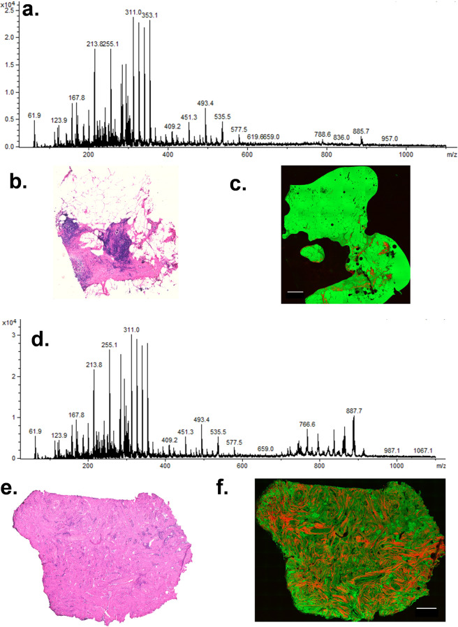
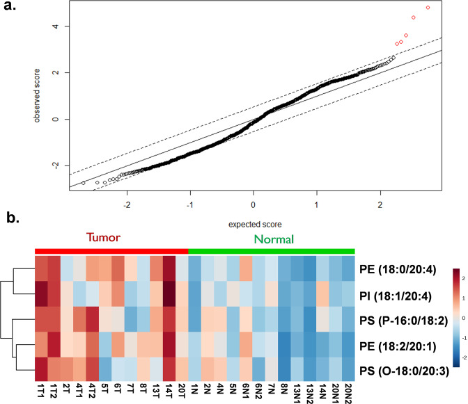
Optimal resection of breast tumors requires removing cancer with a rim of normal tissue while preserving uninvolved regions of the breast. Surgical and pathological techniques that permit rapid molecular characterization of tissue could facilitate such resections. Mass spectrometry (MS) is increasingly used in the research setting to detect and classify tumors and has the potential to detect cancer at surgical margins. Here, we describe the ex vivo intraoperative clinical application of MS using a liquid micro-junction surface sample probe (LMJ-SSP) to assess breast cancer margins. In a midpoint analysis of a registered clinical trial, surgical specimens from 21 women with treatment naïve invasive breast cancer were prospectively collected and analyzed at the time of surgery with subsequent histopathological determination. Normal and tumor breast specimens from the lumpectomy resected by the surgeon were smeared onto glass slides for rapid analysis. Lipidomic profiles were acquired from these specimens using LMJ-SSP MS in negative ionization mode within the operating suite and post-surgery analysis of the data revealed five candidate ions separating tumor from healthy tissue in this limited dataset. More data is required before considering the ions as candidate markers. Here, we present an application of ambient MS within the operating room to analyze breast cancer tissue and surgical margins. Lessons learned from these initial promising studies are being used to further evaluate the five candidate biomarkers and to further refine and optimize intraoperative MS as a tool for surgical guidance in breast cancer.
乳腺肿瘤的最佳切除需要在保留乳腺未受累区域的同时,切除带有正常组织边缘的癌症。能够对组织进行快速分子特征分析的手术和病理技术有助于此类切除。质谱(MS)在研究环境中越来越多地用于检测和分类肿瘤,并且有潜力在手术切缘检测癌症。在此,我们描述了使用液体微连接表面采样探针(LMJ-SSP)进行质谱的离体术中临床应用,以评估乳腺癌切缘。在一项注册临床试验的中期分析中,前瞻性收集了21例未经治疗的浸润性乳腺癌女性的手术标本,并在手术时进行分析,随后进行组织病理学测定。将外科医生切除的肿块切除标本中的正常和肿瘤乳腺标本涂抹在载玻片上进行快速分析。在手术室中使用LMJ-SSP MS以负离子模式从这些标本中获取脂质组学图谱,术后数据分析显示在这个有限的数据集中有五个候选离子可区分肿瘤组织和健康组织。在将这些离子视为候选标志物之前,还需要更多数据。在此,我们展示了在手术室内应用常压质谱分析乳腺癌组织和手术切缘。从这些初步的有前景的研究中吸取的经验教训正用于进一步评估这五个候选生物标志物,并进一步完善和优化术中质谱作为乳腺癌手术指导工具的应用。