Chen Hui-Zhu, Wang Xin-Rong, Zhao Fu-Min, Chen Xi-Jian, Li Xue-Sheng, Ning Gang, Guo Ying-Kun
Department of Radiology, Key Laboratory of Birth Defects and Related Diseases of Women and Children of Ministry of Education, West China Second University Hospital, Sichuan University, Chengdu, China.
PET/MR Department, GE Healthcare, Shanghai, China.
Front Oncol. 2021 Aug 31;11:711648. doi: 10.3389/fonc.2021.711648. eCollection 2021.
To develop and validate a radiomics model for predicting preoperative lymph node (LN) metastasis in high-grade serous ovarian cancer (HGSOC).
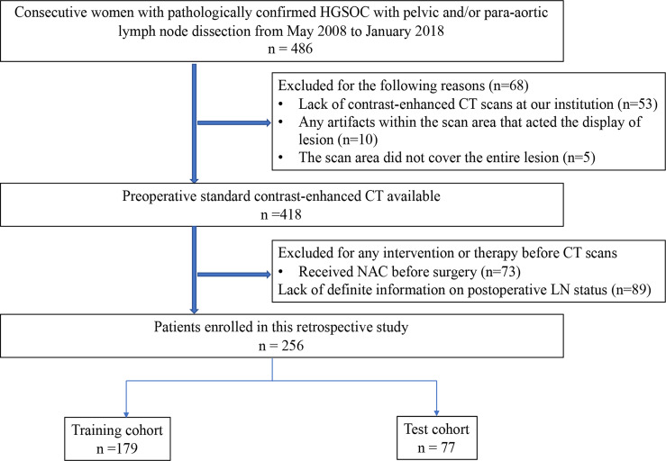
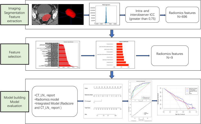
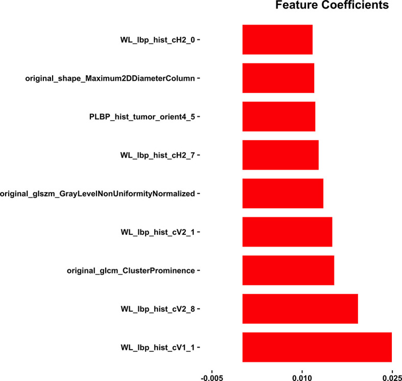
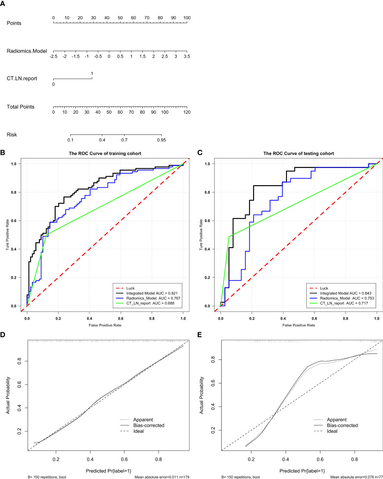
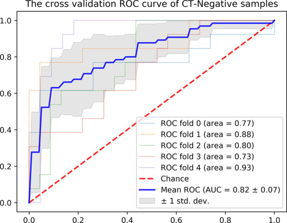
From May 2008 to January 2018, a total of 256 eligible HGSOC patients who underwent tumor resection and LN dissection were divided into a training cohort (n=179) and a test cohort (n=77) in a 7:3 ratio. A Radiomics Model was developed based on a training cohort of 179 patients. A radiomics signature (defined as the Radscore) was selected by using the random forest method. Logistics regression was used as the classifier for modeling. An Integrated Model that incorporated the Radscore and CT_reported LN status (CT_LN_report) was developed and presented as a radiomics nomogram. Its performance was determined by the area under the curve (AUC), calibration, and decision curve. The radiomics nomogram was internally tested in an independent test cohort (n=77) and a CT-LN-report negative subgroup (n=179) using the formula derived from the training cohort.
The AUC value of the CT_LN_report was 0.688 (95% CI: 0.626, 0.759) in the training cohort and 0.717 (95% CI: 0.630, 0.804) in the test cohort. The Radiomics Model yielded an AUC of 0.767 (95% CI: 0.696, 0.837) in the training cohort and 0.753 (95% CI: 0.640, 0.866) in the test. The radiomics nomogram demonstrated favorable calibration and discrimination in the training cohort (AUC=0.821), test cohort (AUC=0.843), and CT-LN-report negative subgroup (AUC=0.82), outperforming the Radiomics Model and CT_LN_report alone.
The radiomics nomogram derived from portal phase CT images performed well in predicting LN metastasis in HGSOC and could be recommended as a new, convenient, and non-invasive method to aid in clinical decision-making.
开发并验证一种用于预测高级别浆液性卵巢癌(HGSOC)术前淋巴结(LN)转移的放射组学模型。
2008年5月至2018年1月,共有256例接受肿瘤切除和LN清扫的符合条件的HGSOC患者按7:3的比例分为训练队列(n = 179)和测试队列(n = 77)。基于179例患者的训练队列开发了放射组学模型。使用随机森林方法选择放射组学特征(定义为Radscore)。采用逻辑回归作为建模的分类器。开发了一个整合Radscore和CT报告的LN状态(CT_LN_report)的整合模型,并将其呈现为放射组学列线图。通过曲线下面积(AUC)、校准和决策曲线来确定其性能。使用从训练队列得出的公式,在独立测试队列(n = 77)和CT-LN报告阴性亚组(n = 179)中对放射组学列线图进行内部测试。
在训练队列中,CT_LN_report的AUC值为0.688(95%CI:0.626,0.759),在测试队列中为0.717(95%CI:0.630,0.804)。放射组学模型在训练队列中的AUC为(0.767)(95%CI:0.696,0.837),在测试中为0.753(95%CI:0.640,0.866)。放射组学列线图在训练队列(AUC = 0.821)、测试队列(AUC = 0.843)和CT-LN报告阴性亚组(AUC = 0.82)中显示出良好的校准和区分能力,优于单独的放射组学模型和CT_LN_report。
源自门静脉期CT图像的放射组学列线图在预测HGSOC中的LN转移方面表现良好,可作为一种新的、方便且无创的方法来辅助临床决策。