• 文献检索
  • 文档翻译
  • 深度研究
  • 学术资讯
  • Suppr Zotero 插件Zotero 插件
  • 邀请有礼
  • 套餐&价格
  • 历史记录
应用&插件
Suppr Zotero 插件Zotero 插件浏览器插件Mac 客户端Windows 客户端微信小程序
定价
高级版会员购买积分包购买API积分包
服务
文献检索文档翻译深度研究API 文档MCP 服务
关于我们
关于 Suppr公司介绍联系我们用户协议隐私条款
关注我们

Suppr 超能文献

核心技术专利:CN118964589B侵权必究
粤ICP备2023148730 号-1Suppr @ 2026

文献检索

告别复杂PubMed语法,用中文像聊天一样搜索,搜遍4000万医学文献。AI智能推荐,让科研检索更轻松。

立即免费搜索

文件翻译

保留排版,准确专业,支持PDF/Word/PPT等文件格式,支持 12+语言互译。

免费翻译文档

深度研究

AI帮你快速写综述,25分钟生成高质量综述,智能提取关键信息,辅助科研写作。

立即免费体验

心肌缺血/再灌注及重塑过程中心肌细胞与成纤维细胞之间的通讯:转化生长因子-β、结缔组织生长因子、肾素血管紧张素轴及非编码RNA分子的作用

Communication Between Cardiomyocytes and Fibroblasts During Cardiac Ischemia/Reperfusion and Remodeling: Roles of TGF-β, CTGF, the Renin Angiotensin Axis, and Non-coding RNA Molecules.

作者信息

Flores-Vergara Raúl, Olmedo Ivonne, Aránguiz Pablo, Riquelme Jaime Andrés, Vivar Raúl, Pedrozo Zully

机构信息

Advanced Center for Chronic Diseases (ACCDiS), Facultad de Ciencias Químicas y Farmacéuticas & Facultad de Medicina, Universidad de Chile, Santiago de Chile, Chile.

Programa de Fisiología y Biofísica, Instituto de Ciencias Biomédicas (ICBM), Facultad de Medicina, Universidad de Chile, Santiago de Chile, Chile.

出版信息

Front Physiol. 2021 Sep 3;12:716721. doi: 10.3389/fphys.2021.716721. eCollection 2021.

DOI:10.3389/fphys.2021.716721
PMID:34539441
原文链接:https://pmc.ncbi.nlm.nih.gov/articles/PMC8446518/
Abstract

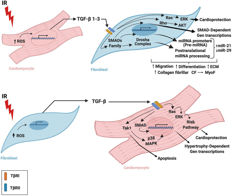
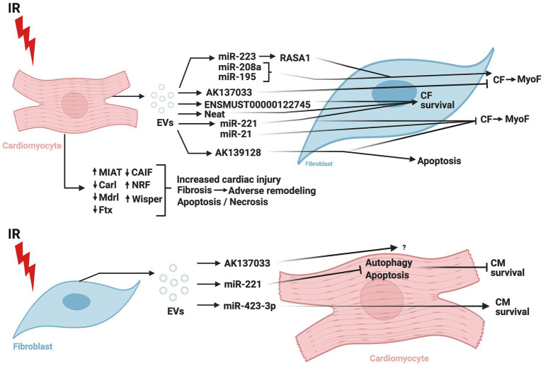
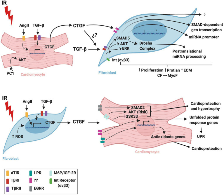
Communication between cells is a foundational concept for understanding the physiology and pathology of biological systems. Paracrine/autocrine signaling, direct cell-to-cell interplay, and extracellular matrix interactions are three types of cell communication that regulate responses to different stimuli. In the heart, cardiomyocytes, fibroblasts, and endothelial cells interact to form the cardiac tissue. Under pathological conditions, such as myocardial infarction, humoral factors released by these cells may induce tissue damage or protection, depending on the type and concentration of molecules secreted. Cardiac remodeling is also mediated by the factors secreted by cardiomyocytes and fibroblasts that are involved in the extensive reciprocal interactions between these cells. Identifying the molecules and cellular signal pathways implicated in these processes will be crucial for creating effective tissue-preserving treatments during or after reperfusion. Numerous therapies to protect cardiac tissue from reperfusion-induced injury have been explored, and ample pre-clinical research has attempted to identify drugs or techniques to mitigate cardiac damage. However, despite great success in animal models, it has not been possible to completely translate these cardioprotective effects to human applications. This review provides a current summary of the principal molecules, pathways, and mechanisms underlying cardiomyocyte and cardiac fibroblast crosstalk during ischemia/reperfusion injury. We also discuss pre-clinical molecules proposed as treatments for myocardial infarction and provide a clinical perspective on these potential therapeutic agents.

摘要

细胞间通讯是理解生物系统生理学和病理学的一个基本概念。旁分泌/自分泌信号传导、细胞间直接相互作用以及细胞外基质相互作用是调节对不同刺激反应的三种细胞通讯类型。在心脏中,心肌细胞、成纤维细胞和内皮细胞相互作用形成心脏组织。在病理条件下,如心肌梗死,这些细胞释放的体液因子可能会根据分泌分子的类型和浓度诱导组织损伤或保护作用。心脏重塑也由心肌细胞和成纤维细胞分泌的因子介导,这些因子参与了这些细胞之间广泛的相互作用。确定这些过程中涉及的分子和细胞信号通路对于在再灌注期间或之后创建有效的组织保护治疗至关重要。已经探索了许多保护心脏组织免受再灌注损伤的疗法,并且大量临床前研究试图确定减轻心脏损伤的药物或技术。然而,尽管在动物模型中取得了巨大成功,但尚未能够将这些心脏保护作用完全转化为人类应用。本综述提供了关于缺血/再灌注损伤期间心肌细胞和心脏成纤维细胞相互作用的主要分子、途径和机制的当前总结。我们还讨论了作为心肌梗死治疗方法提出的临床前分子,并提供了关于这些潜在治疗剂的临床观点。

https://cdn.ncbi.nlm.nih.gov/pmc/blobs/dc92/8446518/be7b6f227da6/fphys-12-716721-g004.jpg
https://cdn.ncbi.nlm.nih.gov/pmc/blobs/dc92/8446518/4e027599008d/fphys-12-716721-g001.jpg
https://cdn.ncbi.nlm.nih.gov/pmc/blobs/dc92/8446518/de22c916061d/fphys-12-716721-g002.jpg
https://cdn.ncbi.nlm.nih.gov/pmc/blobs/dc92/8446518/cd595e70e232/fphys-12-716721-g003.jpg
https://cdn.ncbi.nlm.nih.gov/pmc/blobs/dc92/8446518/be7b6f227da6/fphys-12-716721-g004.jpg

相似文献

1
Communication Between Cardiomyocytes and Fibroblasts During Cardiac Ischemia/Reperfusion and Remodeling: Roles of TGF-β, CTGF, the Renin Angiotensin Axis, and Non-coding RNA Molecules.心肌缺血/再灌注及重塑过程中心肌细胞与成纤维细胞之间的通讯:转化生长因子-β、结缔组织生长因子、肾素血管紧张素轴及非编码RNA分子的作用
Front Physiol. 2021 Sep 3;12:716721. doi: 10.3389/fphys.2021.716721. eCollection 2021.
2
The role of endothelial autocrine NRG1/ERBB4 signaling in cardiac remodeling.内皮细胞自分泌 NRG1/ERBB4 信号在心脏重构中的作用。
Am J Physiol Heart Circ Physiol. 2020 Aug 1;319(2):H443-H455. doi: 10.1152/ajpheart.00176.2020. Epub 2020 Jul 3.
3
The role of angiotensin II, endothelin-1 and transforming growth factor-beta as autocrine/paracrine mediators of stretch-induced cardiomyocyte hypertrophy.血管紧张素II、内皮素-1及转化生长因子-β作为牵张诱导心肌细胞肥大的自分泌/旁分泌介质的作用
Mol Cell Biochem. 2001 Feb;218(1-2):113-24. doi: 10.1023/a:1007279700705.
4
Cardiomyocyte-derived small extracellular vesicles can signal eNOS activation in cardiac microvascular endothelial cells to protect against Ischemia/Reperfusion injury.心肌细胞衍生的小细胞外囊泡可在心脏微血管内皮细胞中发出信号,激活 eNOS,从而防止缺血/再灌注损伤。
Theranostics. 2020 Sep 23;10(25):11754-11774. doi: 10.7150/thno.43163. eCollection 2020.
5
Age-dependent functional crosstalk between cardiac fibroblasts and cardiomyocytes in a 3D engineered cardiac tissue.三维工程化心脏组织中心肌成纤维细胞与心肌细胞之间的年龄依赖性功能串扰
Acta Biomater. 2017 Jun;55:120-130. doi: 10.1016/j.actbio.2017.04.027. Epub 2017 Apr 25.
6
Polycystin-1 mitigates damage and regulates CTGF expression through AKT activation during cardiac ischemia/reperfusion.多囊蛋白-1在心脏缺血/再灌注期间通过激活AKT减轻损伤并调节结缔组织生长因子的表达。
Biochim Biophys Acta Mol Basis Dis. 2021 Jan 1;1867(1):165986. doi: 10.1016/j.bbadis.2020.165986. Epub 2020 Oct 13.
7
The Association of Ascorbic Acid, Deferoxamine and N-Acetylcysteine Improves Cardiac Fibroblast Viability and Cellular Function Associated with Tissue Repair Damaged by Simulated Ischemia/Reperfusion.抗坏血酸、去铁胺和N-乙酰半胱氨酸联合应用可改善心肌成纤维细胞活力及与模拟缺血/再灌注损伤组织修复相关的细胞功能。
Antioxidants (Basel). 2019 Dec 3;8(12):614. doi: 10.3390/antiox8120614.
8
Pleiotropic cardiac functions controlled by ischemia-induced lncRNA H19.缺血诱导的长链非编码 RNA H19 控制的心脏多效性功能。
J Mol Cell Cardiol. 2020 Sep;146:43-59. doi: 10.1016/j.yjmcc.2020.07.001. Epub 2020 Jul 8.
9
CTGF/CCN2 is an autocrine regulator of cardiac fibrosis.CTGF/CCN2 是心脏纤维化的自分泌调节剂。
J Mol Cell Cardiol. 2018 Aug;121:205-211. doi: 10.1016/j.yjmcc.2018.07.130. Epub 2018 Jul 21.
10
TGF-β1 prevents simulated ischemia/reperfusion-induced cardiac fibroblast apoptosis by activation of both canonical and non-canonical signaling pathways.转化生长因子-β1通过激活经典和非经典信号通路来预防模拟缺血/再灌注诱导的心脏成纤维细胞凋亡。
Biochim Biophys Acta. 2013 Jun;1832(6):754-62. doi: 10.1016/j.bbadis.2013.02.004. Epub 2013 Feb 15.

引用本文的文献

1
Stromal-platelet membrane-inspired nanoparticles (SPIN) for targeted heart repair.用于靶向心脏修复的基质血小板膜启发型纳米颗粒(SPIN)
Bioact Mater. 2025 Jul 7;53:45-57. doi: 10.1016/j.bioactmat.2025.06.055. eCollection 2025 Nov.
2
Small extracellular vesicles enhance the survival of Sca-1+ cardiac stem cells against ROS-induced ischemic-reoxygenation injury in vitro.小细胞外囊泡可增强Sca-1+心脏干细胞在体外抵抗活性氧诱导的缺血-再灌注损伤的存活能力。
Biol Res. 2025 Mar 5;58(1):12. doi: 10.1186/s40659-025-00593-7.
3
Cardiomyocyte-restricted MIAT deletion is sufficient to protect against murine myocardial infarction.

本文引用的文献

1
Long non-coding RNAs: From disease code to drug role.长链非编码RNA:从疾病密码到药物作用
Acta Pharm Sin B. 2021 Feb;11(2):340-354. doi: 10.1016/j.apsb.2020.10.001. Epub 2020 Oct 10.
2
Therapeutic advances of miRNAs: A preclinical and clinical update.微小RNA的治疗进展:临床前与临床最新情况
J Adv Res. 2020 Aug 29;28:127-138. doi: 10.1016/j.jare.2020.08.012. eCollection 2021 Feb.
3
Polycystin-1 mitigates damage and regulates CTGF expression through AKT activation during cardiac ischemia/reperfusion.多囊蛋白-1在心脏缺血/再灌注期间通过激活AKT减轻损伤并调节结缔组织生长因子的表达。
心肌细胞特异性MIAT缺失足以预防小鼠心肌梗死。
Cell Death Discov. 2025 Feb 20;11(1):70. doi: 10.1038/s41420-025-02352-9.
4
Serpina3k lactylation protects from cardiac ischemia reperfusion injury.丝氨酸蛋白酶抑制剂A3K(Serpina3k)乳酸化可保护心脏免受缺血再灌注损伤。
Nat Commun. 2025 Jan 25;16(1):1012. doi: 10.1038/s41467-024-55589-w.
5
Hyperuricemia suppresses lumican, exacerbating adverse remodeling after myocardial infarction by promoting fibroblast phenotype transition.高尿酸血症抑制了 lumican,通过促进成纤维细胞表型转变,加剧心肌梗死后的不良重构。
J Transl Med. 2024 Oct 31;22(1):983. doi: 10.1186/s12967-024-05778-4.
6
Ferroptosis in diabetic cardiomyopathy: Advances in cardiac fibroblast-cardiomyocyte interactions.糖尿病性心肌病中的铁死亡:心脏成纤维细胞与心肌细胞相互作用的研究进展
Heliyon. 2024 Jul 28;10(15):e35219. doi: 10.1016/j.heliyon.2024.e35219. eCollection 2024 Aug 15.
7
Cardiac Fibroblasts Enhance MMP2 Activity to Suppress Gap Junction Function in Cardiomyocytes.心脏成纤维细胞增强MMP2活性以抑制心肌细胞中的缝隙连接功能。
Appl Biochem Biotechnol. 2024 Dec;196(12):8493-8512. doi: 10.1007/s12010-024-04986-4. Epub 2024 Jun 15.
8
Modelling the pathology and treatment of cardiac fibrosis in vascularised atrial and ventricular cardiac microtissues.构建血管化心房和心室心脏微组织中心脏纤维化的病理模型及治疗方法。
Front Cardiovasc Med. 2023 Sep 1;10:1156759. doi: 10.3389/fcvm.2023.1156759. eCollection 2023.
9
Sevoflurane Improves Ventricular Conduction by Exosomes Derived from Rat Cardiac Fibroblasts After Hypothermic Global Ischemia-Reperfusion Injury.七氟醚通过低温全球缺血再灌注损伤后大鼠心肌成纤维细胞来源的外泌体改善心室传导。
Drug Des Devel Ther. 2023 Jun 12;17:1719-1732. doi: 10.2147/DDDT.S408595. eCollection 2023.
10
Cardiomyocyte-targeting exosomes from sulforaphane-treated fibroblasts affords cardioprotection in infarcted rats.经萝卜硫素处理的成纤维细胞来源的心肌细胞靶向细胞外囊泡为梗死大鼠提供心脏保护。
J Transl Med. 2023 May 9;21(1):313. doi: 10.1186/s12967-023-04155-x.
Biochim Biophys Acta Mol Basis Dis. 2021 Jan 1;1867(1):165986. doi: 10.1016/j.bbadis.2020.165986. Epub 2020 Oct 13.
4
Crosstalk between cardiomyocytes and noncardiomyocytes is essential to prevent cardiomyocyte apoptosis induced by proteasome inhibition.心肌细胞和非心肌细胞之间的串扰对于预防蛋白酶体抑制诱导的心肌细胞凋亡至关重要。
Cell Death Dis. 2020 Sep 19;11(9):783. doi: 10.1038/s41419-020-03005-8.
5
Position-specific oxidation of miR-1 encodes cardiac hypertrophy.miR-1的位点特异性氧化编码心肌肥大。
Nature. 2020 Aug;584(7820):279-285. doi: 10.1038/s41586-020-2586-0. Epub 2020 Aug 5.
6
An overview of the classical and tissue-derived renin-angiotensin-aldosterone system and its genetic polymorphisms in essential hypertension.经典和组织衍生的肾素-血管紧张素-醛固酮系统概述及其在原发性高血压中的遗传多态性。
Steroids. 2020 Nov;163:108701. doi: 10.1016/j.steroids.2020.108701. Epub 2020 Jul 24.
7
Adverse Cardiac Remodelling after Acute Myocardial Infarction: Old and New Biomarkers.急性心肌梗死后的心脏不良重构:新旧生物标志物。
Dis Markers. 2020 Jun 12;2020:1215802. doi: 10.1155/2020/1215802. eCollection 2020.
8
Targeting JMJD3 histone demethylase mediates cardiac fibrosis and cardiac function following myocardial infarction.靶向 JMJD3 组蛋白去甲基化酶可减轻心肌梗死后的心脏纤维化和心功能障碍。
Biochem Biophys Res Commun. 2020 Aug 6;528(4):671-677. doi: 10.1016/j.bbrc.2020.05.115. Epub 2020 Jun 6.
9
Exosomal lncRNA AK139128 Derived from Hypoxic Cardiomyocytes Promotes Apoptosis and Inhibits Cell Proliferation in Cardiac Fibroblasts.低氧心肌细胞来源的外泌体 lncRNA AK139128 促进心肌成纤维细胞凋亡和抑制细胞增殖。
Int J Nanomedicine. 2020 May 12;15:3363-3376. doi: 10.2147/IJN.S240660. eCollection 2020.
10
Inhibition of chymotrypsin-like activity of the proteasome by ixazomib prevents mitochondrial dysfunction during myocardial ischemia.依沙佐米抑制蛋白酶体糜蛋白酶样活性可防止心肌缺血期间的线粒体功能障碍。
PLoS One. 2020 May 26;15(5):e0233591. doi: 10.1371/journal.pone.0233591. eCollection 2020.