• 文献检索
  • 文档翻译
  • 深度研究
  • 学术资讯
  • Suppr Zotero 插件Zotero 插件
  • 邀请有礼
  • 套餐&价格
  • 历史记录
应用&插件
Suppr Zotero 插件Zotero 插件浏览器插件Mac 客户端Windows 客户端微信小程序
定价
高级版会员购买积分包购买API积分包
服务
文献检索文档翻译深度研究API 文档MCP 服务
关于我们
关于 Suppr公司介绍联系我们用户协议隐私条款
关注我们

Suppr 超能文献

核心技术专利:CN118964589B侵权必究
粤ICP备2023148730 号-1Suppr @ 2026

文献检索

告别复杂PubMed语法,用中文像聊天一样搜索,搜遍4000万医学文献。AI智能推荐,让科研检索更轻松。

立即免费搜索

文件翻译

保留排版,准确专业,支持PDF/Word/PPT等文件格式,支持 12+语言互译。

免费翻译文档

深度研究

AI帮你快速写综述,25分钟生成高质量综述,智能提取关键信息,辅助科研写作。

立即免费体验

眼组织高维分析的成像质谱流式技术

Imaging mass cytometry for high-dimensional tissue profiling in the eye.

机构信息

Faculty of Medicine, Eye Center, University of Freiburg, Killianstrasse 5, 79106, Freiburg, Germany.

Institute of Anatomy, Wuerzburg University, Wuerzburg, Germany.

出版信息

BMC Ophthalmol. 2021 Sep 20;21(1):338. doi: 10.1186/s12886-021-02099-8.

DOI:10.1186/s12886-021-02099-8
PMID:34544377
原文链接:https://pmc.ncbi.nlm.nih.gov/articles/PMC8454101/
Abstract

BACKGROUND

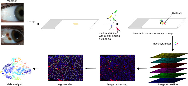
Imaging mass cytometry (IMC) combines the principles of flow cytometry and mass spectrometry (MS) with laser scanning spatial resolution and offers unique advantages for the analysis of tissue samples in unprecedented detail. In contrast to conventional immunohistochemistry, which is limited in its application by the number of possible fluorochrome combinations, IMC uses isoptope-coupled antibodies that allow multiplex analysis of up to 40 markers in the same tissue section simultaneously.

METHODS

In this report we use IMC to analyze formalin-fixed, paraffin-embedded conjunctival tissue. We performed a 18-biomarkers IMC analysis of conjunctival tissue to determine and summarize the possibilities, relevance and limitations of IMC for deciphering the biology and pathology of ocular diseases.

RESULTS

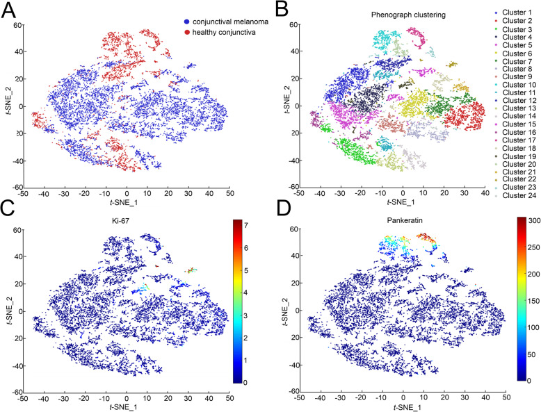
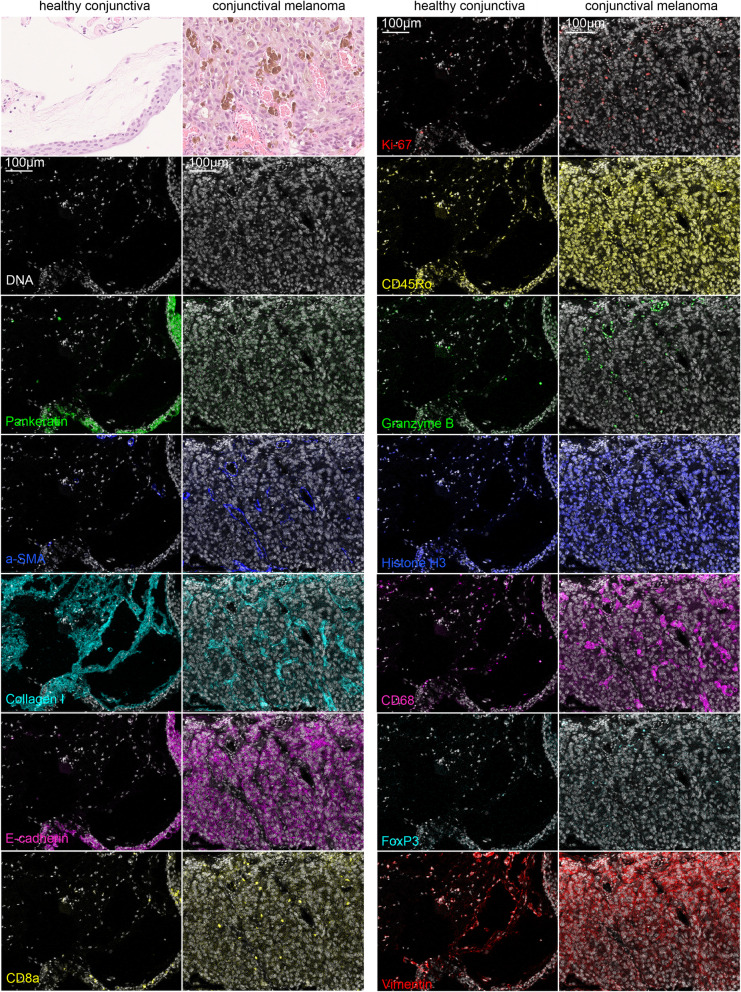
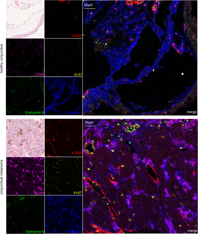
Without modifying the manufacturer's protocol, we observed positive and plausible staining for 12 of 18 biomarkers. Subsequent bioinformatical single-cell analysis and phenograph clustering identified 24 different cellular clusters with distinct expression profiles with respect to the markers used.

CONCLUSIONS

IMC enables highly multiplexed imaging of ocular samples at subcellular resolution. IMC is an innovative and feasible method, providing new insights into ocular disease pathogenesis that will be valuable for basic research, drug discovery and clinical diagnostics.

摘要

背景

成像质谱流式细胞术(IMC)结合了流式细胞术和质谱(MS)的原理,具有激光扫描空间分辨率,为以前所未有的细节分析组织样本提供了独特的优势。与传统的免疫组织化学相比,免疫组织化学受到可能的荧光染料组合数量的限制,而 IMC 则使用等光耦连抗体,允许在同一切片上同时对多达 40 个标记物进行多重分析。

方法

在本报告中,我们使用 IMC 分析了福尔马林固定、石蜡包埋的结膜组织。我们对结膜组织进行了 18 种生物标志物的 IMC 分析,以确定和总结 IMC 用于破译眼部疾病生物学和病理学的可能性、相关性和局限性。

结果

在不修改制造商方案的情况下,我们观察到 18 种生物标志物中的 12 种具有阳性和合理的染色。随后的单细胞生物信息学分析和表型聚类确定了 24 个不同的细胞簇,这些细胞簇在使用的标记物方面具有不同的表达谱。

结论

IMC 能够以亚细胞分辨率对眼部样本进行高度多重成像。IMC 是一种创新且可行的方法,为眼部疾病发病机制提供了新的见解,这将对基础研究、药物发现和临床诊断具有重要价值。

https://cdn.ncbi.nlm.nih.gov/pmc/blobs/3f6e/8454101/4644994c0238/12886_2021_2099_Fig4_HTML.jpg
https://cdn.ncbi.nlm.nih.gov/pmc/blobs/3f6e/8454101/a283f87dea7e/12886_2021_2099_Fig1_HTML.jpg
https://cdn.ncbi.nlm.nih.gov/pmc/blobs/3f6e/8454101/dac6b80a5786/12886_2021_2099_Fig2_HTML.jpg
https://cdn.ncbi.nlm.nih.gov/pmc/blobs/3f6e/8454101/f21b2b314b3f/12886_2021_2099_Fig3_HTML.jpg
https://cdn.ncbi.nlm.nih.gov/pmc/blobs/3f6e/8454101/4644994c0238/12886_2021_2099_Fig4_HTML.jpg

相似文献

1
Imaging mass cytometry for high-dimensional tissue profiling in the eye.眼组织高维分析的成像质谱流式技术
BMC Ophthalmol. 2021 Sep 20;21(1):338. doi: 10.1186/s12886-021-02099-8.
2
Research Techniques Made Simple: Use of Imaging Mass Cytometry for Dermatological Research and Clinical Applications.研究技巧简单化:利用影像质谱细胞仪进行皮肤科研究和临床应用。
J Invest Dermatol. 2021 Apr;141(4):705-712.e1. doi: 10.1016/j.jid.2020.12.008.
3
MATISSE: a method for improved single cell segmentation in imaging mass cytometry.MATISSE:一种用于成像质谱细胞术中单细胞分割改进的方法。
BMC Biol. 2021 May 11;19(1):99. doi: 10.1186/s12915-021-01043-y.
4
Coupling imaging mass cytometry with Alcian blue histochemical staining for a single-slide approach.结合成像质谱流式细胞术和阿尔辛蓝组织化学染色的单幻灯片方法。
Front Immunol. 2024 Apr 29;15:1379154. doi: 10.3389/fimmu.2024.1379154. eCollection 2024.
5
Imaging Mass Cytometry in Immuno-Oncology.免疫肿瘤学中的成像质谱流式细胞术
Methods Mol Biol. 2023;2614:1-15. doi: 10.1007/978-1-0716-2914-7_1.
6
Method to Investigate the Distribution of Water-Soluble Drug-Delivery Systems in Fresh Frozen Tissues Using Imaging Mass Cytometry.利用成像质谱细胞术研究水溶性药物递送系统在新鲜冷冻组织中的分布。
Anal Chem. 2021 Mar 2;93(8):3742-3749. doi: 10.1021/acs.analchem.0c03908. Epub 2021 Feb 19.
7
Imaging Mass Cytometry.成像质谱流式细胞术
Cytometry A. 2017 Feb;91(2):160-169. doi: 10.1002/cyto.a.23053. Epub 2017 Feb 3.
8
Biomarker Discovery in Patients with Immunotherapy-Treated Melanoma with Imaging Mass Cytometry.应用影像质谱细胞术发现免疫治疗黑色素瘤患者的生物标志物。
Clin Cancer Res. 2021 Apr 1;27(7):1987-1996. doi: 10.1158/1078-0432.CCR-20-3340. Epub 2021 Jan 27.
9
Research Techniques Made Simple: Experimental Methodology for Imaging Mass Cytometry.研究技巧轻松学:影像质谱细胞术的实验方法。
J Invest Dermatol. 2021 Mar;141(3):467-473.e1. doi: 10.1016/j.jid.2020.11.022.
10
DNA-barcoded signal amplification for imaging mass cytometry enables sensitive and highly multiplexed tissue imaging.DNA 条码信号放大用于成像质谱流式细胞术,实现了灵敏和高多重化的组织成像。
Nat Methods. 2023 Sep;20(9):1304-1309. doi: 10.1038/s41592-023-01976-y. Epub 2023 Aug 31.

引用本文的文献

1
Evolutionary and immune microenvironment dynamics during neoadjuvant treatment of esophageal adenocarcinoma.食管腺癌新辅助治疗期间的进化与免疫微环境动态变化
Nat Cancer. 2025 May 14. doi: 10.1038/s43018-025-00955-w.
2
Immune Cytolytic Activity and Strategies for Therapeutic Treatment.免疫细胞溶解活性与治疗策略。
Int J Mol Sci. 2024 Mar 23;25(7):3624. doi: 10.3390/ijms25073624.
3
Hyalocytes-guardians of the vitreoretinal interface.玻璃体细胞——视网膜界面的守护者。

本文引用的文献

1
Transcript levels of keratin 1/5/6/14/15/16/17 as potential prognostic indicators in melanoma patients.角蛋白 1/5/6/14/15/16/17 的转录水平作为黑色素瘤患者潜在的预后指标。
Sci Rep. 2021 Jan 13;11(1):1023. doi: 10.1038/s41598-020-80336-8.
2
PD-L1 Expression in 65 Conjunctival Melanomas and Its Association with Clinical Outcome.65例结膜黑色素瘤中程序性死亡受体配体1(PD-L1)的表达及其与临床结局的关联
Int J Mol Sci. 2020 Nov 30;21(23):9147. doi: 10.3390/ijms21239147.
3
Conjunctival nevi and melanoma: multiparametric immunohistochemical analysis, including p16, SOX10, HMB45, and Ki-67.
Graefes Arch Clin Exp Ophthalmol. 2024 Sep;262(9):2765-2784. doi: 10.1007/s00417-024-06448-3. Epub 2024 Apr 3.
4
Dual-modality imaging of immunofluorescence and imaging mass cytometry for whole-slide imaging and accurate segmentation.免疫荧光双模态成像和成像质谱流式细胞术的全切片成像和精确分割。
Cell Rep Methods. 2023 Oct 23;3(10):100595. doi: 10.1016/j.crmeth.2023.100595. Epub 2023 Sep 22.
5
Naproxen chemoprevention induces proliferation of cytotoxic lymphocytes in Lynch Syndrome colorectal mucosa.萘普生化学预防诱导林奇综合征结直肠黏膜中细胞毒性淋巴细胞的增殖。
Front Immunol. 2023 May 3;14:1162669. doi: 10.3389/fimmu.2023.1162669. eCollection 2023.
6
The promise and challenge of spatial omics in dissecting tumour microenvironment and the role of AI.空间组学在剖析肿瘤微环境中的前景与挑战以及人工智能的作用
Front Oncol. 2023 May 1;13:1172314. doi: 10.3389/fonc.2023.1172314. eCollection 2023.
7
Dual-modality imaging of immunofluorescence and imaging mass cytometry for whole slide imaging with accurate single-cell segmentation.用于全玻片成像并具备精确单细胞分割功能的免疫荧光和成像质谱流式细胞术的双模态成像。
bioRxiv. 2023 Feb 23:2023.02.23.529718. doi: 10.1101/2023.02.23.529718.
8
Hyalocyte origin, structure, and imaging.玻璃体细胞的起源、结构与成像。
Expert Rev Ophthalmol. 2022;17(4):233-248. doi: 10.1080/17469899.2022.2100762. Epub 2022 Sep 6.
9
Single-Cell Protein and Transcriptional Characterization of Epiretinal Membranes From Patients With Proliferative Vitreoretinopathy.单细胞蛋白质组学和增生性玻璃体视网膜病变患者眼内膜的转录特征分析。
Invest Ophthalmol Vis Sci. 2022 May 2;63(5):17. doi: 10.1167/iovs.63.5.17.
10
In-Depth Molecular Characterization of Neovascular Membranes Suggests a Role for Hyalocyte-to-Myofibroblast Transdifferentiation in Proliferative Diabetic Retinopathy.深入的分子特征分析表明,玻璃体细胞向肌成纤维细胞转分化在增生性糖尿病视网膜病变中起作用。
Front Immunol. 2021 Nov 2;12:757607. doi: 10.3389/fimmu.2021.757607. eCollection 2021.
结膜痣和黑色素瘤:包括 p16、SOX10、HMB45 和 Ki-67 的多参数免疫组织化学分析。
Hum Pathol. 2020 Sep;103:107-119. doi: 10.1016/j.humpath.2020.07.020. Epub 2020 Jul 21.
4
Immunohistochemical analysis of benign and malignant melanocytic lesions of the conjunctiva using double-staining.应用双重染色法对结膜的良性和恶性黑色素细胞病变进行免疫组织化学分析。
Can J Ophthalmol. 2019 Dec;54(6):699-707. doi: 10.1016/j.jcjo.2019.03.015. Epub 2019 Jun 14.
5
Mass Cytometry Imaging for the Study of Human Diseases-Applications and Data Analysis Strategies.质谱细胞术成像在人类疾病研究中的应用及数据分析策略。
Front Immunol. 2019 Nov 14;10:2657. doi: 10.3389/fimmu.2019.02657. eCollection 2019.
6
A 40-Marker Panel for High Dimensional Characterization of Cancer Immune Microenvironments by Imaging Mass Cytometry.通过影像质谱流式细胞术对癌症免疫微环境进行高维特征分析的 40 标志物面板
Front Immunol. 2019 Oct 29;10:2534. doi: 10.3389/fimmu.2019.02534. eCollection 2019.
7
A Map of Human Type 1 Diabetes Progression by Imaging Mass Cytometry.基于影像质谱流式细胞术的人类 1 型糖尿病进展图谱
Cell Metab. 2019 Mar 5;29(3):755-768.e5. doi: 10.1016/j.cmet.2018.11.014. Epub 2019 Jan 31.
8
Mass Spectrometry Imaging and Integration with Other Imaging Modalities for Greater Molecular Understanding of Biological Tissues.质谱成像与其他成像模式的整合,以增进对生物组织的分子理解。
Mol Imaging Biol. 2018 Dec;20(6):888-901. doi: 10.1007/s11307-018-1267-y.
9
Simultaneous Multiplexed Imaging of mRNA and Proteins with Subcellular Resolution in Breast Cancer Tissue Samples by Mass Cytometry.利用质谱流式细胞术对乳腺癌组织样本进行亚细胞分辨率的 mRNA 和蛋白质的同时多重成像。
Cell Syst. 2018 Jan 24;6(1):25-36.e5. doi: 10.1016/j.cels.2017.12.001. Epub 2017 Dec 27.
10
Organizational hierarchy and structural diversity of microvascular pericytes in adult mouse cortex.成年小鼠皮层中小血管周细胞的组织层次结构和结构多样性。
J Cereb Blood Flow Metab. 2019 Mar;39(3):411-425. doi: 10.1177/0271678X17732229. Epub 2017 Sep 21.