• 文献检索
  • 文档翻译
  • 深度研究
  • 学术资讯
  • Suppr Zotero 插件Zotero 插件
  • 邀请有礼
  • 套餐&价格
  • 历史记录
应用&插件
Suppr Zotero 插件Zotero 插件浏览器插件Mac 客户端Windows 客户端微信小程序
定价
高级版会员购买积分包购买API积分包
服务
文献检索文档翻译深度研究API 文档MCP 服务
关于我们
关于 Suppr公司介绍联系我们用户协议隐私条款
关注我们

Suppr 超能文献

核心技术专利:CN118964589B侵权必究
粤ICP备2023148730 号-1Suppr @ 2026

文献检索

告别复杂PubMed语法,用中文像聊天一样搜索,搜遍4000万医学文献。AI智能推荐,让科研检索更轻松。

立即免费搜索

文件翻译

保留排版,准确专业,支持PDF/Word/PPT等文件格式,支持 12+语言互译。

免费翻译文档

深度研究

AI帮你快速写综述,25分钟生成高质量综述,智能提取关键信息,辅助科研写作。

立即免费体验

高级影像学和计算技术在恶性脑胶质瘤的诊断和预后评估中的应用。

Advanced Imaging and Computational Techniques for the Diagnostic and Prognostic Assessment of Malignant Gliomas.

机构信息

From the Division of Neuroradiology, Department of Radiology.

Department of Computational Medicine and Bioinformatics, Michigan Medicine.

出版信息

Cancer J. 2021;27(5):344-352. doi: 10.1097/PPO.0000000000000545.

DOI:10.1097/PPO.0000000000000545
PMID:34570448
原文链接:https://pmc.ncbi.nlm.nih.gov/articles/PMC8480516/
Abstract

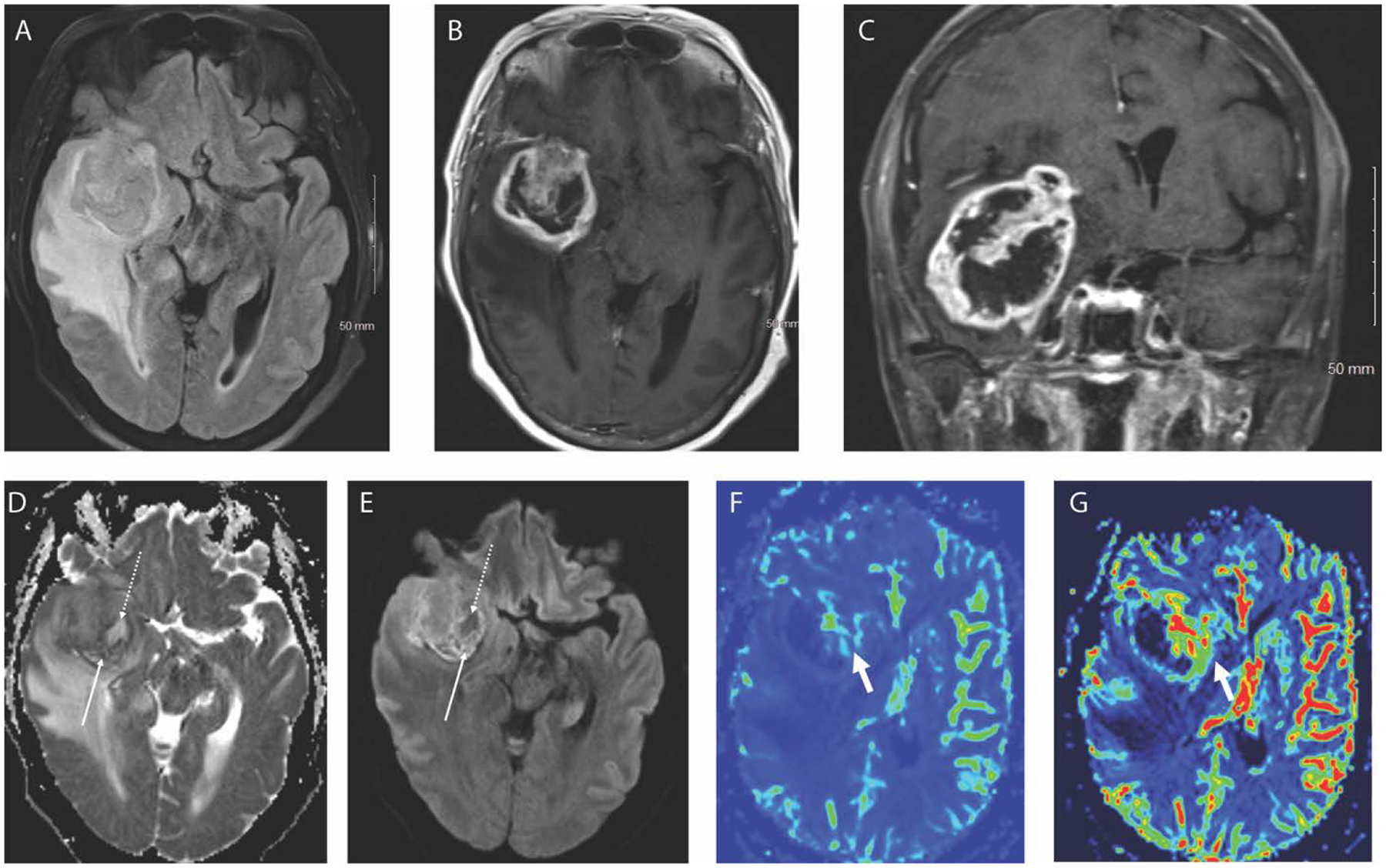
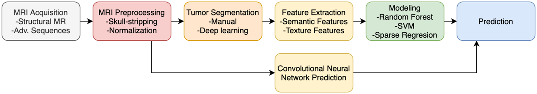
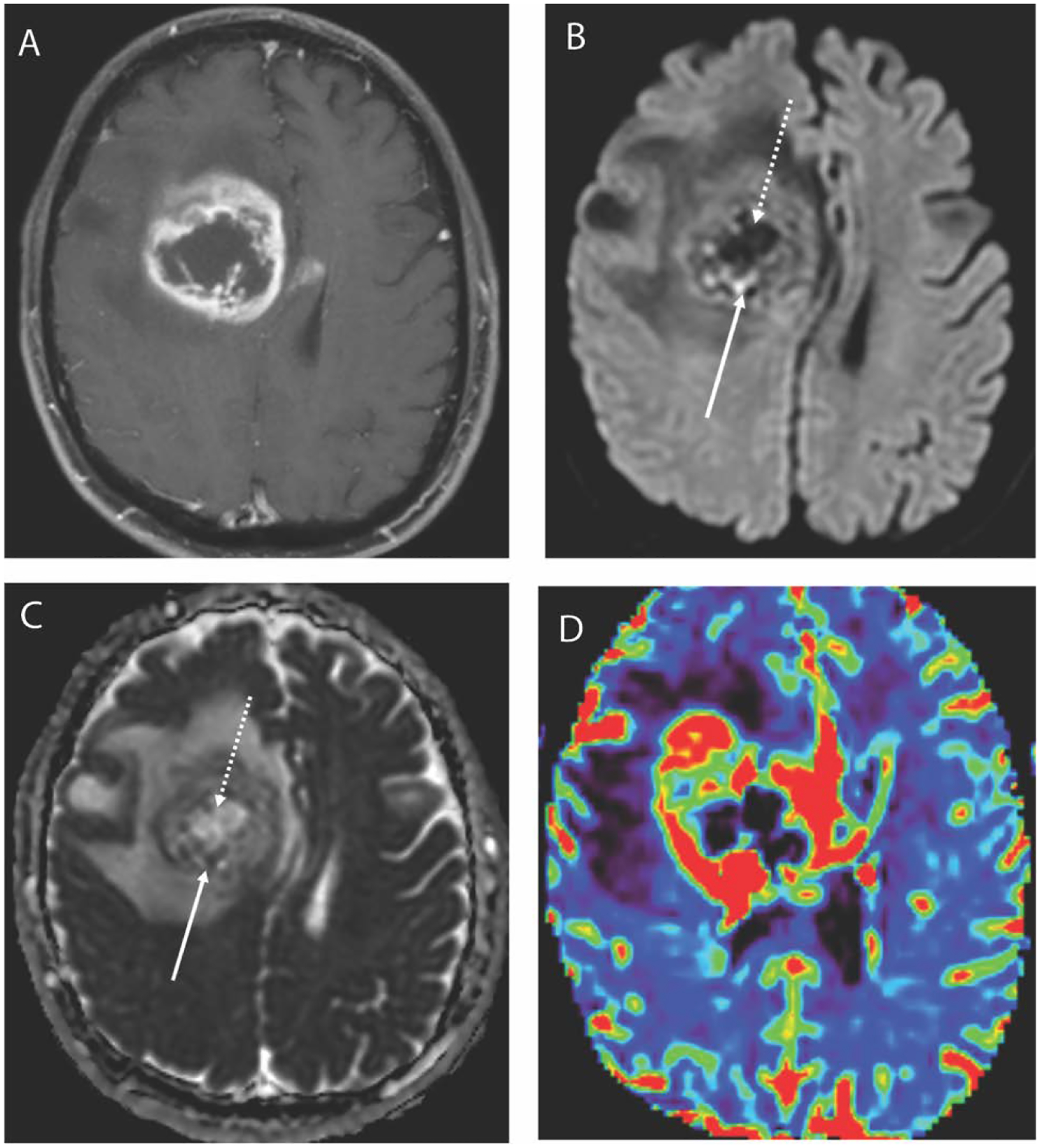
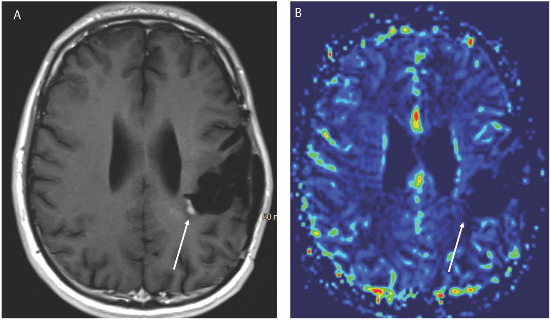
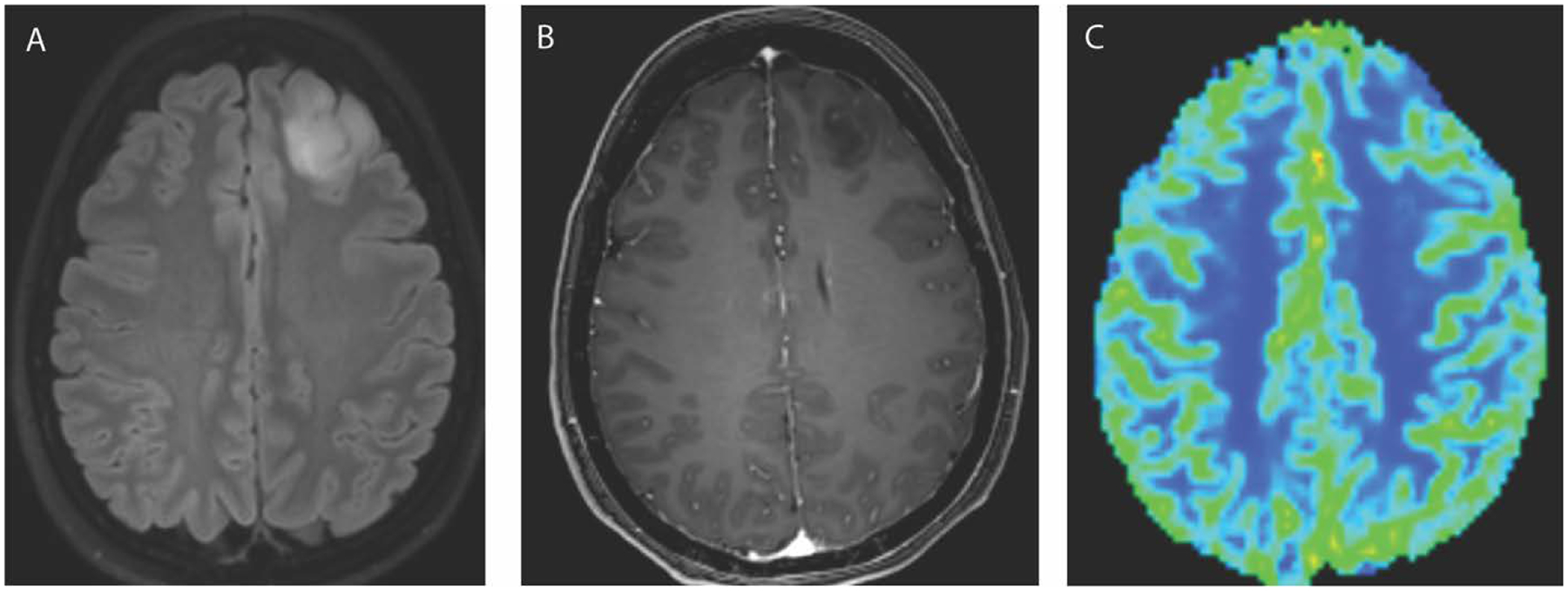
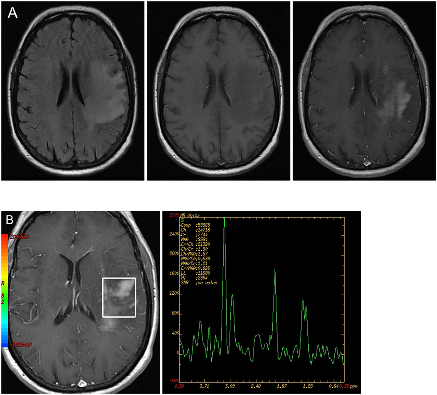
Advanced imaging techniques provide a powerful tool to assess the intratumoral and intertumoral heterogeneity of gliomas. Advances in the molecular understanding of glioma subgroups may allow improved diagnostic assessment combining imaging and molecular tumor features, with enhanced prognostic utility and implications for patient treatment. In this article, a comprehensive overview of the physiologic basis for conventional and advanced imaging techniques is presented, and clinical applications before and after treatment are discussed. An introduction to the principles of radiomics and the advanced integration of imaging, clinical outcomes, and genomic data highlights the future potential for this field of research to better stratify and select patients for standard as well as investigational therapies.

摘要

高级成像技术为评估胶质瘤的瘤内和瘤间异质性提供了有力的工具。对胶质瘤亚群分子认识的进步可能允许通过将成像和分子肿瘤特征相结合来进行改进的诊断评估,从而提高预后能力并影响患者的治疗。本文全面介绍了常规和高级成像技术的生理基础,并讨论了治疗前后的临床应用。对放射组学原理以及成像、临床结果和基因组数据的高级整合的介绍突出了该研究领域的未来潜力,可以更好地对患者进行分层并选择标准治疗和研究性治疗。

https://cdn.ncbi.nlm.nih.gov/pmc/blobs/dfa9/8480516/4f24b1668544/nihms-1731563-f0006.jpg
https://cdn.ncbi.nlm.nih.gov/pmc/blobs/dfa9/8480516/e80fa7bb7ace/nihms-1731563-f0001.jpg
https://cdn.ncbi.nlm.nih.gov/pmc/blobs/dfa9/8480516/6f7e5e2340e2/nihms-1731563-f0002.jpg
https://cdn.ncbi.nlm.nih.gov/pmc/blobs/dfa9/8480516/f8a4d5eb952d/nihms-1731563-f0003.jpg
https://cdn.ncbi.nlm.nih.gov/pmc/blobs/dfa9/8480516/81fed00783dd/nihms-1731563-f0004.jpg
https://cdn.ncbi.nlm.nih.gov/pmc/blobs/dfa9/8480516/93dcfcc0db04/nihms-1731563-f0005.jpg
https://cdn.ncbi.nlm.nih.gov/pmc/blobs/dfa9/8480516/4f24b1668544/nihms-1731563-f0006.jpg

相似文献

1
Advanced Imaging and Computational Techniques for the Diagnostic and Prognostic Assessment of Malignant Gliomas.高级影像学和计算技术在恶性脑胶质瘤的诊断和预后评估中的应用。
Cancer J. 2021;27(5):344-352. doi: 10.1097/PPO.0000000000000545.
2
Imaging of intratumoral heterogeneity in high-grade glioma.高级别胶质瘤瘤内异质性的影像学表现。
Cancer Lett. 2020 May 1;477:97-106. doi: 10.1016/j.canlet.2020.02.025. Epub 2020 Feb 27.
3
Adult Glioma WHO Classification Update, Genomics, and Imaging: What the Radiologists Need to Know.成人胶质瘤的世界卫生组织分类更新、基因组学与影像学:放射科医生需要了解的内容。
Top Magn Reson Imaging. 2020 Apr;29(2):71-82. doi: 10.1097/RMR.0000000000000234.
4
Radiomics in gliomas: clinical implications of computational modeling and fractal-based analysis.脑胶质瘤的放射组学:基于计算模型和分形分析的临床意义。
Neuroradiology. 2020 Jul;62(7):771-790. doi: 10.1007/s00234-020-02403-1. Epub 2020 Apr 6.
5
Radiogenomics of Gliomas.脑胶质瘤的放射组学
Radiol Clin North Am. 2021 May;59(3):441-455. doi: 10.1016/j.rcl.2021.02.002.
6
Machine learning and radiomic phenotyping of lower grade gliomas: improving survival prediction.机器学习和低级别胶质瘤的放射组学表型:改善生存预测。
Eur Radiol. 2020 Jul;30(7):3834-3842. doi: 10.1007/s00330-020-06737-5. Epub 2020 Mar 11.
7
Imaging Correlates of Adult Glioma Genotypes.成人脑胶质瘤基因型的影像学相关性。
Radiology. 2017 Aug;284(2):316-331. doi: 10.1148/radiol.2017151930.
8
Reverse Engineering Glioma Radiomics to Conventional Neuroimaging.将胶质瘤放射组学逆向工程到传统神经影像学
Neurol Med Chir (Tokyo). 2021 Sep 15;61(9):505-514. doi: 10.2176/nmc.ra.2021-0133. Epub 2021 Aug 6.
9
Radiomics risk score may be a potential imaging biomarker for predicting survival in isocitrate dehydrogenase wild-type lower-grade gliomas.放射组学风险评分可能是预测异柠檬酸脱氢酶野生型低级别胶质瘤患者生存的潜在影像生物标志物。
Eur Radiol. 2020 Dec;30(12):6464-6474. doi: 10.1007/s00330-020-07089-w. Epub 2020 Aug 1.
10
Machine Learning for the Prediction of Molecular Markers in Glioma on Magnetic Resonance Imaging: A Systematic Review and Meta-Analysis.基于磁共振成像的机器学习预测脑胶质瘤分子标志物的系统评价和荟萃分析。
Neurosurgery. 2021 Jun 15;89(1):31-44. doi: 10.1093/neuros/nyab103.

引用本文的文献

1
A Systematic Review of the Current Status and Quality of Radiomics for Glioma Differential Diagnosis.胶质瘤鉴别诊断中影像组学现状与质量的系统评价
Cancers (Basel). 2022 May 31;14(11):2731. doi: 10.3390/cancers14112731.

本文引用的文献

1
Radiomics and radiogenomics in gliomas: a contemporary update.脑胶质瘤的影像组学和放射组学:当代进展。
Br J Cancer. 2021 Aug;125(5):641-657. doi: 10.1038/s41416-021-01387-w. Epub 2021 May 6.
2
Clinical Applications of Magnetic Resonance Spectroscopy in Brain Tumors: From Diagnosis to Treatment.磁共振波谱在脑肿瘤中的临床应用:从诊断到治疗。
Radiol Clin North Am. 2021 May;59(3):349-362. doi: 10.1016/j.rcl.2021.01.004. Epub 2021 Mar 23.
3
Application of Diffusion Weighted Imaging and Diffusion Tensor Imaging in the Pretreatment and Post-treatment of Brain Tumor.
弥散加权成像和弥散张量成像在脑肿瘤治疗前后的应用。
Radiol Clin North Am. 2021 May;59(3):335-347. doi: 10.1016/j.rcl.2021.01.003. Epub 2021 Mar 23.
4
Imaging Findings of New Entities and Patterns in Brain Tumor: Isocitrate Dehydrogenase Mutant, Isocitrate Dehydrogenase Wild-Type, Codeletion, and MGMT Methylation.脑肿瘤新实体和模式的影像学表现:异柠檬酸脱氢酶突变型、异柠檬酸脱氢酶野生型、缺失型和 MGMT 甲基化。
Radiol Clin North Am. 2021 May;59(3):305-322. doi: 10.1016/j.rcl.2021.01.001. Epub 2021 Mar 24.
5
Advanced MR techniques in glioblastoma imaging-upcoming challenges and how to face them.高级磁共振技术在脑胶质瘤成像中的应用——即将面临的挑战及应对策略。
Eur Radiol. 2021 Sep;31(9):6652-6654. doi: 10.1007/s00330-021-07978-8. Epub 2021 Apr 22.
6
Low conductivity on electrical properties tomography demonstrates unique tumor habitats indicating progression in glioblastoma.电特性断层成像中的低传导性表明胶质母细胞瘤的进展存在独特的肿瘤生境。
Eur Radiol. 2021 Sep;31(9):6655-6665. doi: 10.1007/s00330-021-07976-w. Epub 2021 Apr 20.
7
The cIMPACT-NOW updates and their significance to current neuro-oncology practice.cIMPACT-NOW更新及其对当前神经肿瘤学实践的意义。
Neurooncol Pract. 2020 Aug 29;8(1):4-10. doi: 10.1093/nop/npaa055. eCollection 2021 Feb.
8
Mapping tumour heterogeneity with pulsed 3D CEST MRI in non-enhancing glioma at 3 T.在 3T 下使用脉冲 3D CEST MRI 对非增强性脑胶质瘤进行肿瘤异质性成像。
MAGMA. 2022 Feb;35(1):53-62. doi: 10.1007/s10334-021-00911-6. Epub 2021 Feb 19.
9
Evaluation of perfusion MRI value for tumor progression assessment after glioma radiotherapy: A systematic review and meta-analysis.评估灌注 MRI 对胶质瘤放疗后肿瘤进展评估的价值:系统评价和荟萃分析。
Medicine (Baltimore). 2020 Dec 24;99(52):e23766. doi: 10.1097/MD.0000000000023766.
10
Identifying the spatial and temporal dynamics of molecularly-distinct glioblastoma sub-populations.鉴定分子异质性胶质母细胞瘤亚群的时空动态。
Math Biosci Eng. 2020 Jul 16;17(5):4905-4941. doi: 10.3934/mbe.2020267.