Division of Cancer Genetics, Chiba Cancer Center Research Institute, Nitona, Chuoh-ku, Chiba, Japan.
BMC Mol Cell Biol. 2021 Oct 7;22(1):52. doi: 10.1186/s12860-021-00391-5.
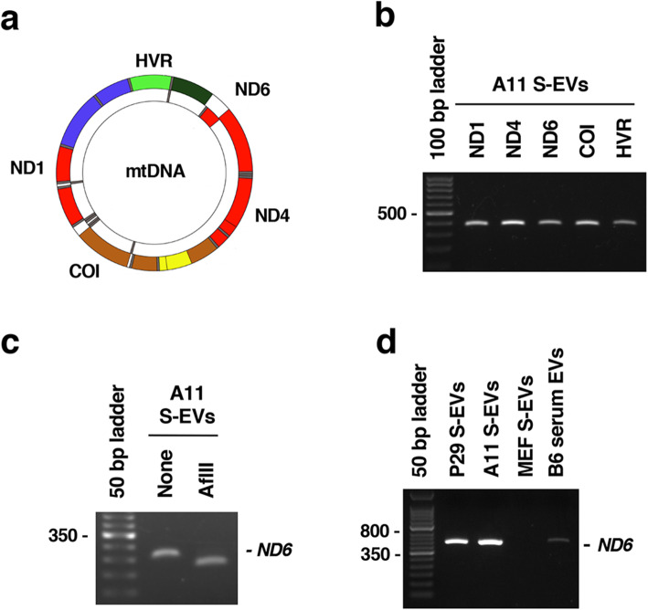
Mitochondrial DNA (mtDNA) carrying certain pathogenic mutations or single nucleotide variants (SNVs) enhances the invasion and metastasis of tumor cells, and some of these mutations are homoplasmic in tumor cells and even in tumor tissues. On the other hand, intercellular transfer of mitochondria and cellular components via extracellular vesicles (EVs) and tunneling nanotubes (TNTs) has recently attracted intense attention in terms of cell-to-cell communication in the tumor microenvironment. It remains unclear whether metastasis-enhancing pathogenic mutant mtDNA in tumor cells is intercellularly transferred between tumor cells and stromal cells. In this study, we investigated whether mtDNA with the NADH dehydrogenase subunit 6 (ND6) G13997A pathogenic mutation in highly metastatic cells can be horizontally transferred to low-metastatic cells and stromal cells in the tumor microenvironment.
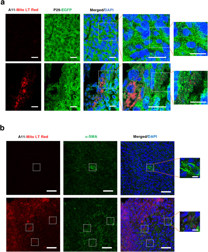
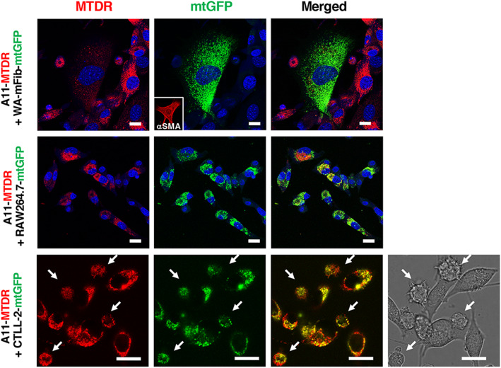
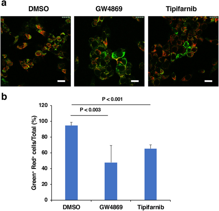
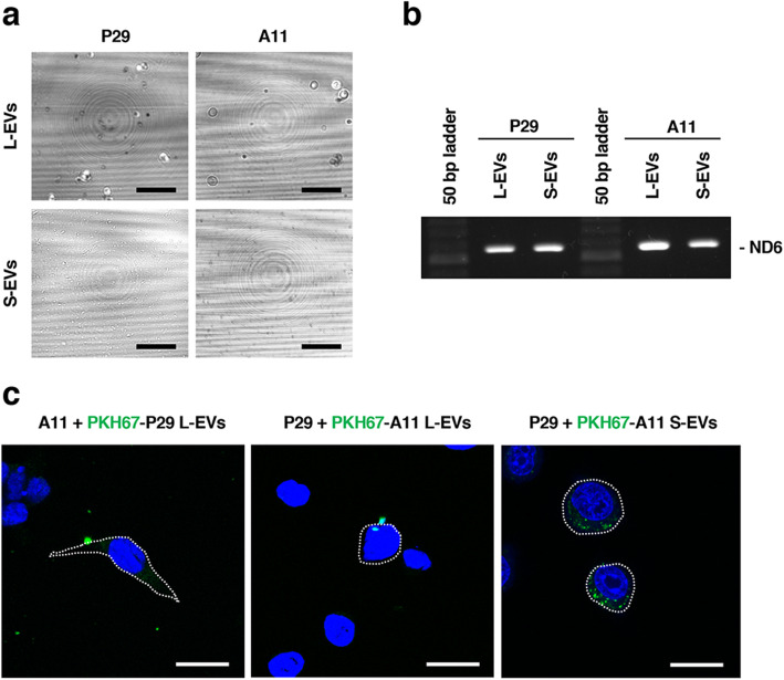
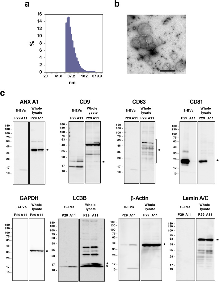
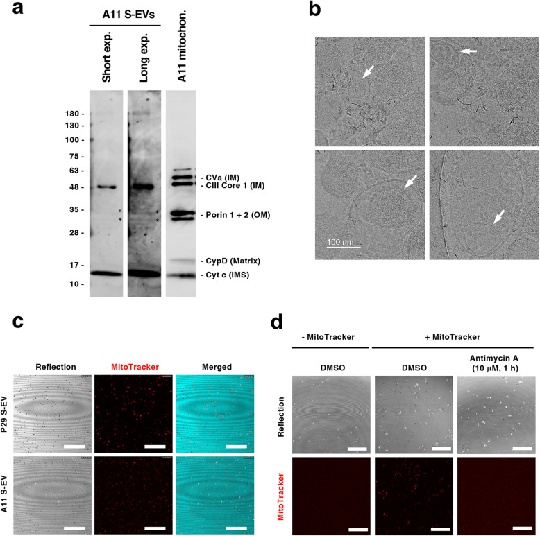
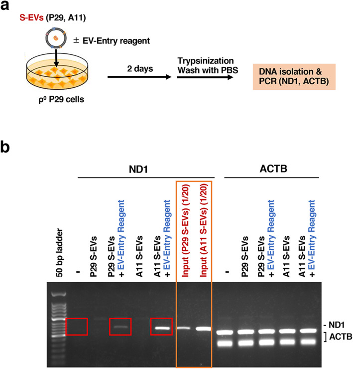
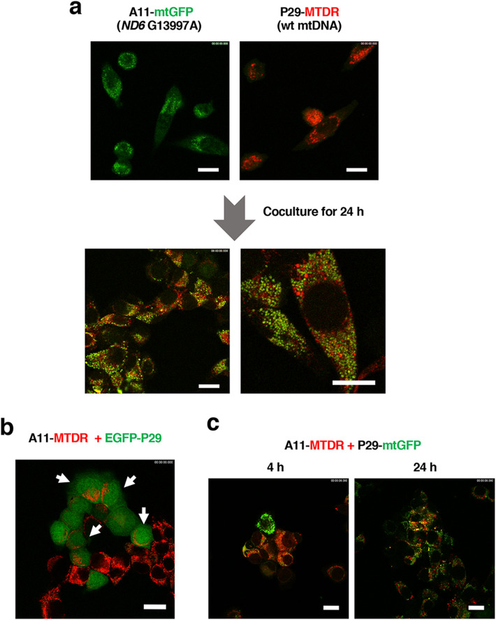
When MitoTracker Deep Red-labeled high-metastatic Lewis lung carcinoma A11 cells carrying the ND6 G13997A mtDNA mutation were cocultured with CellLight mitochondria-GFP-labeled low-metastatic P29 cells harboring wild-type mtDNA, bidirectional transfer of red- and green-colored vesicles, probably mitochondria-related EVs, was observed in a time-dependent manner. Similarly, intercellular transfer of mitochondria-related EVs occurred between A11 cells and α-smooth muscle actin (α-SMA)-positive cancer-associated fibroblasts (CAFs, WA-mFib), macrophages (RAW264.7) and cytotoxic T cells (CTLL-2). Intercellular transfer was suppressed by inhibitors of EV release. The large and small EV fractions (L-EV and S-EV, respectively) prepared from the conditioned medium by differential ultracentrifugation both were found to contain mtDNA, although only S-EVs were efficiently incorporated into the cells. Several subpopulations had evidence of LC3-II and contained degenerated mitochondrial components in the S-EV fraction, signaling to the existence of autophagy-related S-EVs. Interestingly, the S-EV fraction contained a MitoTracker-positive subpopulation, which was inhibited by the respiration inhibitor antimycin A, indicating the presence of mitochondria with membrane potential. It was also demonstrated that mtDNA was transferred into mtDNA-less ρ cells after coculture with the S-EV fraction. In syngeneic mouse subcutaneous tumors formed by a mixture of A11 and P29 cells, the mitochondria-related EVs released from A11 cells reached distantly positioned P29 cells and CAFs.
These results suggest that metastasis-enhancing pathogenic mtDNA derived from metastatic tumor cells is transferred to low-metastatic tumor cells and stromal cells via S-EVs in vitro and in the tumor microenvironment, inferring a novel mechanism of enhancement of metastatic potential during tumor progression.
携带有特定致病性突变或单核苷酸变异(SNV)的线粒体 DNA(mtDNA)会增强肿瘤细胞的侵袭和转移能力,其中一些突变在肿瘤细胞中甚至在肿瘤组织中都是同质的。另一方面,通过细胞外囊泡(EVs)和隧道纳米管(TNTs)进行的细胞间线粒体和细胞成分的转移,最近在肿瘤微环境中的细胞间通讯方面引起了强烈关注。目前尚不清楚肿瘤细胞中增强转移的致病性突变 mtDNA 是否在肿瘤细胞与基质细胞之间进行细胞间转移。在这项研究中,我们研究了高度转移性 Lewis 肺癌 A11 细胞中的 NADH 脱氢酶亚单位 6(ND6)G13997A 致病性突变 mtDNA 是否可以水平转移到肿瘤微环境中低转移性的 P29 细胞和基质细胞中。
当用 MitoTracker Deep Red 标记携带有 ND6 G13997A mtDNA 突变的高转移性 Lewis 肺癌 A11 细胞与携带野生型 mtDNA 的 CellLight 线粒体-GFP 标记的低转移性 P29 细胞共培养时,红色和绿色囊泡(可能是与线粒体相关的 EVs)的双向转移在时间依赖性的方式中观察到。同样,与α-平滑肌肌动蛋白(α-SMA)阳性癌症相关成纤维细胞(CAFs,WA-mFib)、巨噬细胞(RAW264.7)和细胞毒性 T 细胞(CTLL-2)之间也发生了与线粒体相关的 EV 细胞间转移。EV 释放抑制剂抑制了细胞间转移。通过差速超速离心从条件培养基中制备的大、小 EV 部分(分别为 L-EV 和 S-EV)均含有 mtDNA,尽管只有 S-EV 能够有效地进入细胞。S-EV 部分中的几个亚群存在 LC3-II 的证据,并含有退化的线粒体成分,表明存在自噬相关的 S-EV。有趣的是,S-EV 部分含有 MitoTracker 阳性亚群,该亚群被呼吸抑制剂安密妥 A 抑制,表明存在具有膜电位的线粒体。还证明,在用 S-EV 部分共培养后,mtDNA 转移到 mtDNA 缺失的 ρ 细胞中。在由 A11 和 P29 细胞混合物形成的同基因小鼠皮下肿瘤中,从 A11 细胞释放的与线粒体相关的 EV 到达远处位置的 P29 细胞和 CAFs。
这些结果表明,源自转移性肿瘤细胞的增强转移的致病性 mtDNA 通过 S-EV 在体外和肿瘤微环境中转移到低转移性肿瘤细胞和基质细胞中,推断出肿瘤进展过程中增强转移潜能的一种新机制。