• 文献检索
  • 文档翻译
  • 深度研究
  • 学术资讯
  • Suppr Zotero 插件Zotero 插件
  • 邀请有礼
  • 套餐&价格
  • 历史记录
应用&插件
Suppr Zotero 插件Zotero 插件浏览器插件Mac 客户端Windows 客户端微信小程序
定价
高级版会员购买积分包购买API积分包
服务
文献检索文档翻译深度研究API 文档MCP 服务
关于我们
关于 Suppr公司介绍联系我们用户协议隐私条款
关注我们

Suppr 超能文献

核心技术专利:CN118964589B侵权必究
粤ICP备2023148730 号-1Suppr @ 2026

文献检索

告别复杂PubMed语法,用中文像聊天一样搜索,搜遍4000万医学文献。AI智能推荐,让科研检索更轻松。

立即免费搜索

文件翻译

保留排版,准确专业,支持PDF/Word/PPT等文件格式,支持 12+语言互译。

免费翻译文档

深度研究

AI帮你快速写综述,25分钟生成高质量综述,智能提取关键信息,辅助科研写作。

立即免费体验

体干涉格子光片成像。

Volumetric interferometric lattice light-sheet imaging.

机构信息

Structural Biology Program, Memorial Sloan Kettering Cancer Center, New York, NY, USA.

出版信息

Nat Biotechnol. 2021 Nov;39(11):1385-1393. doi: 10.1038/s41587-021-01042-y. Epub 2021 Oct 11.

DOI:10.1038/s41587-021-01042-y
PMID:34635835
原文链接:https://pmc.ncbi.nlm.nih.gov/articles/PMC8595582/
Abstract

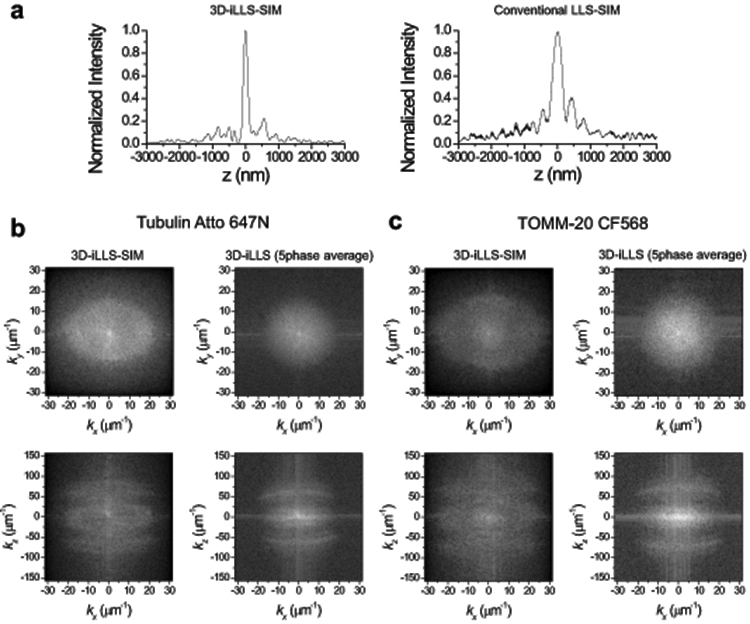
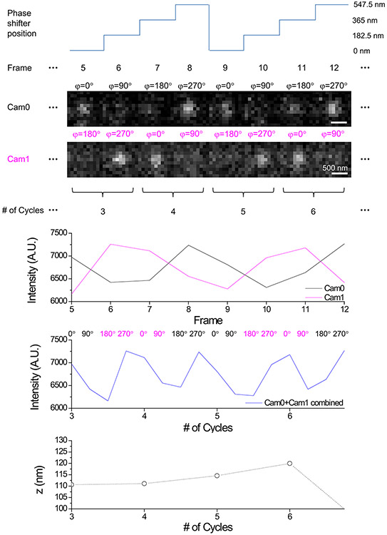
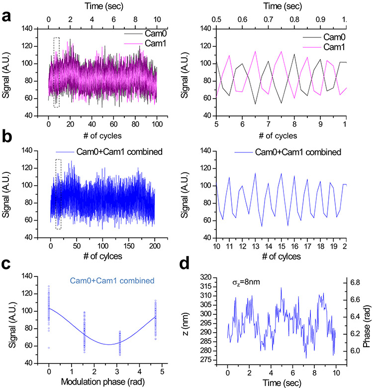
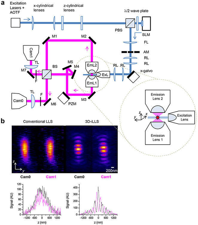
Live cell imaging with high spatiotemporal resolution and high detection sensitivity facilitates the study of the dynamics of cellular structure and function. However, extracting high-resolution 4D (3D space plus time) information from live cells remains challenging, because current methods are slow, require high peak excitation intensities or suffer from high out-of-focus background. Here we present 3D interferometric lattice light-sheet (3D-iLLS) imaging, a technique that requires low excitation light levels and provides high background suppression and substantially improved volumetric resolution by combining 4Pi interferometry with selective plane illumination. We demonstrate that 3D-iLLS has an axial resolution and single-particle localization precision of 100 nm (FWHM) and <10 nm (1σ), respectively. We illustrate the performance of 3D-iLLS in a range of systems: single messenger RNA molecules, nanoscale assemblies of transcription regulators in the nucleus, the microtubule cytoskeleton and mitochondria organelles. The enhanced 4D resolution and increased signal-to-noise ratio of 3D-iLLS will facilitate the analysis of biological processes at the sub-cellular level.

摘要

活细胞的高时空分辨率和高检测灵敏度成像有助于研究细胞结构和功能的动力学。然而,从活细胞中提取高分辨率的 4D(3D 空间加时间)信息仍然具有挑战性,因为目前的方法速度较慢,需要高的峰值激发强度,或者受到高离焦背景的影响。在这里,我们提出了三维干涉晶格光片(3D-iLLS)成像技术,该技术通过将 4Pi 干涉仪与选择性平面照明相结合,在低激发光水平下提供高背景抑制和大大提高的体积分辨率。我们证明了 3D-iLLS 的轴向分辨率和单粒子定位精度分别为 100nm(FWHM)和<10nm(1σ)。我们在一系列系统中说明了 3D-iLLS 的性能:单个信使 RNA 分子、核内转录调控因子的纳米级组装、微管细胞骨架和线粒体细胞器。3D-iLLS 的增强的 4D 分辨率和增加的信噪比将有助于在亚细胞水平分析生物过程。

https://cdn.ncbi.nlm.nih.gov/pmc/blobs/435b/8595582/572a1068884f/nihms-1729175-f0006.jpg
https://cdn.ncbi.nlm.nih.gov/pmc/blobs/435b/8595582/83129ab7e9da/nihms-1729175-f0007.jpg
https://cdn.ncbi.nlm.nih.gov/pmc/blobs/435b/8595582/9aa93554adf9/nihms-1729175-f0008.jpg
https://cdn.ncbi.nlm.nih.gov/pmc/blobs/435b/8595582/724dcbd0af2e/nihms-1729175-f0009.jpg
https://cdn.ncbi.nlm.nih.gov/pmc/blobs/435b/8595582/7717800bafb9/nihms-1729175-f0010.jpg
https://cdn.ncbi.nlm.nih.gov/pmc/blobs/435b/8595582/00cb20bb3f5c/nihms-1729175-f0011.jpg
https://cdn.ncbi.nlm.nih.gov/pmc/blobs/435b/8595582/f2eb10e8ef17/nihms-1729175-f0012.jpg
https://cdn.ncbi.nlm.nih.gov/pmc/blobs/435b/8595582/e65a2e2b64a8/nihms-1729175-f0013.jpg
https://cdn.ncbi.nlm.nih.gov/pmc/blobs/435b/8595582/b174431f8b1f/nihms-1729175-f0014.jpg
https://cdn.ncbi.nlm.nih.gov/pmc/blobs/435b/8595582/fe9193e73a17/nihms-1729175-f0015.jpg
https://cdn.ncbi.nlm.nih.gov/pmc/blobs/435b/8595582/65dfb3e0f58f/nihms-1729175-f0016.jpg
https://cdn.ncbi.nlm.nih.gov/pmc/blobs/435b/8595582/43bcffd40142/nihms-1729175-f0001.jpg
https://cdn.ncbi.nlm.nih.gov/pmc/blobs/435b/8595582/8072b38390a8/nihms-1729175-f0002.jpg
https://cdn.ncbi.nlm.nih.gov/pmc/blobs/435b/8595582/ac8bf214db4f/nihms-1729175-f0003.jpg
https://cdn.ncbi.nlm.nih.gov/pmc/blobs/435b/8595582/c1647d2dc6b0/nihms-1729175-f0004.jpg
https://cdn.ncbi.nlm.nih.gov/pmc/blobs/435b/8595582/6ab327a70f55/nihms-1729175-f0005.jpg
https://cdn.ncbi.nlm.nih.gov/pmc/blobs/435b/8595582/572a1068884f/nihms-1729175-f0006.jpg

相似文献

1
Volumetric interferometric lattice light-sheet imaging.体干涉格子光片成像。
Nat Biotechnol. 2021 Nov;39(11):1385-1393. doi: 10.1038/s41587-021-01042-y. Epub 2021 Oct 11.
2
Resolution doubling in light-sheet microscopy via oblique plane structured illumination.利用斜平面结构照明实现光片显微镜的分辨率倍增。
Nat Methods. 2022 Nov;19(11):1419-1426. doi: 10.1038/s41592-022-01635-8. Epub 2022 Oct 24.
3
Rapid high resolution 3D imaging of expanded biological specimens with lattice light sheet microscopy.晶格层光片显微镜快速高分辨率 3D 成像扩展生物样本。
Methods. 2020 Mar 1;174:11-19. doi: 10.1016/j.ymeth.2019.04.006. Epub 2019 Apr 9.
4
Three-dimensional super-resolution imaging of live whole cells using galvanometer-based structured illumination microscopy.使用基于振镜的结构照明显微镜对活的完整细胞进行三维超分辨率成像。
Opt Express. 2019 Mar 4;27(5):7237-7248. doi: 10.1364/OE.27.007237.
5
Light sheet approaches for improved precision in 3D localization-based super-resolution imaging in mammalian cells [Invited].用于提高哺乳动物细胞中基于三维定位的超分辨率成像精度的光片方法[特邀]
Opt Express. 2018 May 14;26(10):13122-13147. doi: 10.1364/OE.26.013122.
6
Interferometric fluorescent super-resolution microscopy resolves 3D cellular ultrastructure.干涉荧光超分辨率显微镜可解析三维细胞超微结构。
Proc Natl Acad Sci U S A. 2009 Mar 3;106(9):3125-30. doi: 10.1073/pnas.0813131106. Epub 2009 Feb 6.
7
Nanoscale three-dimensional single particle tracking by light-sheet-based double-helix point spread function microscopy.基于光片的双螺旋点扩散函数显微镜的纳米级三维单粒子追踪
Appl Opt. 2016 Jan 20;55(3):449-53. doi: 10.1364/AO.55.000449.
8
Video-rate 3D imaging of living cells using Fourier view-channel-depth light field microscopy.使用傅里叶视通道深度光场显微镜对活细胞进行视频速率的 3D 成像。
Commun Biol. 2023 Dec 12;6(1):1259. doi: 10.1038/s42003-023-05636-x.
9
Three-dimensional structured illumination microscopy with enhanced axial resolution.三维结构光照明显微镜,轴向分辨率增强。
Nat Biotechnol. 2023 Sep;41(9):1307-1319. doi: 10.1038/s41587-022-01651-1. Epub 2023 Jan 26.
10
3D single-molecule super-resolution microscopy with a tilted light sheet.倾斜光片的 3D 单分子超分辨率显微镜。
Nat Commun. 2018 Jan 9;9(1):123. doi: 10.1038/s41467-017-02563-4.

引用本文的文献

1
ClearScope: a fully integrated light sheet theta microscope for sub-cellular resolution imaging without lateral size constraints.ClearScope:一种完全集成的光片θ显微镜,用于亚细胞分辨率成像,且无横向尺寸限制。
bioRxiv. 2024 Aug 19:2024.08.15.608141. doi: 10.1101/2024.08.15.608141.
2
Image restoration of degraded time-lapse microscopy data mediated by near-infrared imaging.近红外成像介导的退化延时显微镜数据的图像恢复。
Nat Methods. 2024 Feb;21(2):311-321. doi: 10.1038/s41592-023-02127-z. Epub 2024 Jan 4.
3
Integrative approaches to study enhancer-promoter communication.
综合方法研究增强子-启动子通讯。
Curr Opin Genet Dev. 2023 Jun;80:102052. doi: 10.1016/j.gde.2023.102052. Epub 2023 May 29.
4
Light-sheets and smart microscopy, an exciting future is dawning.光片和智能显微镜,一个激动人心的未来正在到来。
Commun Biol. 2023 May 9;6(1):502. doi: 10.1038/s42003-023-04857-4.
5
Nanoscale nuclear environments, fine-scale 3D genome organization and transcription regulation.纳米级核环境、精细尺度三维基因组组织与转录调控。
Curr Opin Syst Biol. 2022 Sep;31. doi: 10.1016/j.coisb.2022.100436. Epub 2022 Oct 21.
6
Sidelobe suppression in structured light sheet fluorescence microscopy by the superposition of two light sheets.通过两个光片叠加实现结构光片荧光显微镜中的旁瓣抑制
Biomed Opt Express. 2023 Feb 17;14(3):1178-1191. doi: 10.1364/BOE.481508. eCollection 2023 Mar 1.
7
Three-dimensional structured illumination microscopy with enhanced axial resolution.三维结构光照明显微镜,轴向分辨率增强。
Nat Biotechnol. 2023 Sep;41(9):1307-1319. doi: 10.1038/s41587-022-01651-1. Epub 2023 Jan 26.
8
Understanding immune signaling using advanced imaging techniques.利用先进的成像技术来理解免疫信号。
Biochem Soc Trans. 2022 Apr 29;50(2):853-866. doi: 10.1042/BST20210479.
9
Deciphering the Retinal Epigenome during Development, Disease and Reprogramming: Advancements, Challenges and Perspectives.解析发育、疾病和重编程过程中的视网膜表观基因组:进展、挑战与展望。
Cells. 2022 Feb 25;11(5):806. doi: 10.3390/cells11050806.
10
New insights into promoter-enhancer communication mechanisms revealed by dynamic single-molecule imaging.动态单分子成像揭示的启动子-增强子通讯机制的新见解。
Biochem Soc Trans. 2021 Jun 30;49(3):1299-1309. doi: 10.1042/BST20200963.