Department of Biology, Trinity University, San Antonio, TX, United States.
Front Neural Circuits. 2021 Oct 4;15:731333. doi: 10.3389/fncir.2021.731333. eCollection 2021.
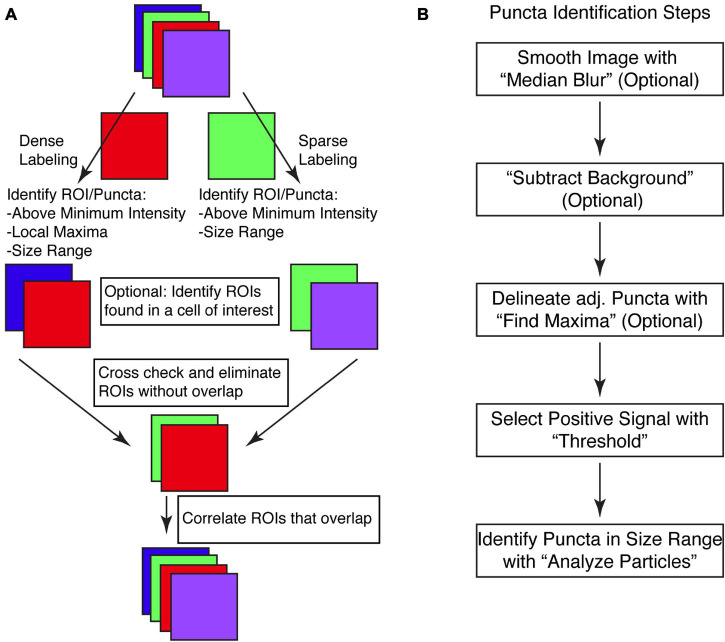
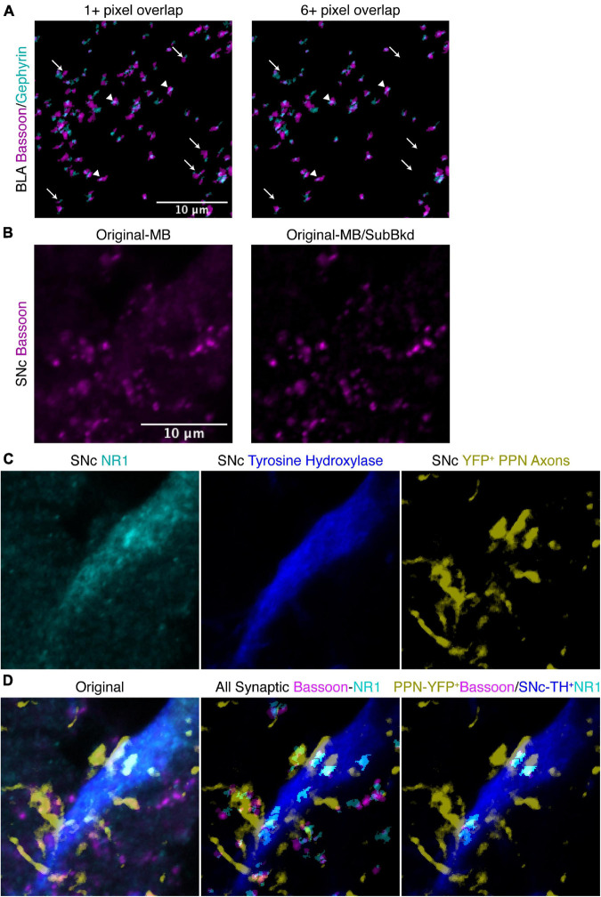
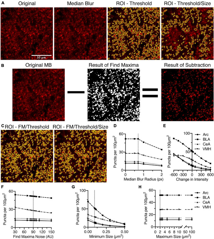
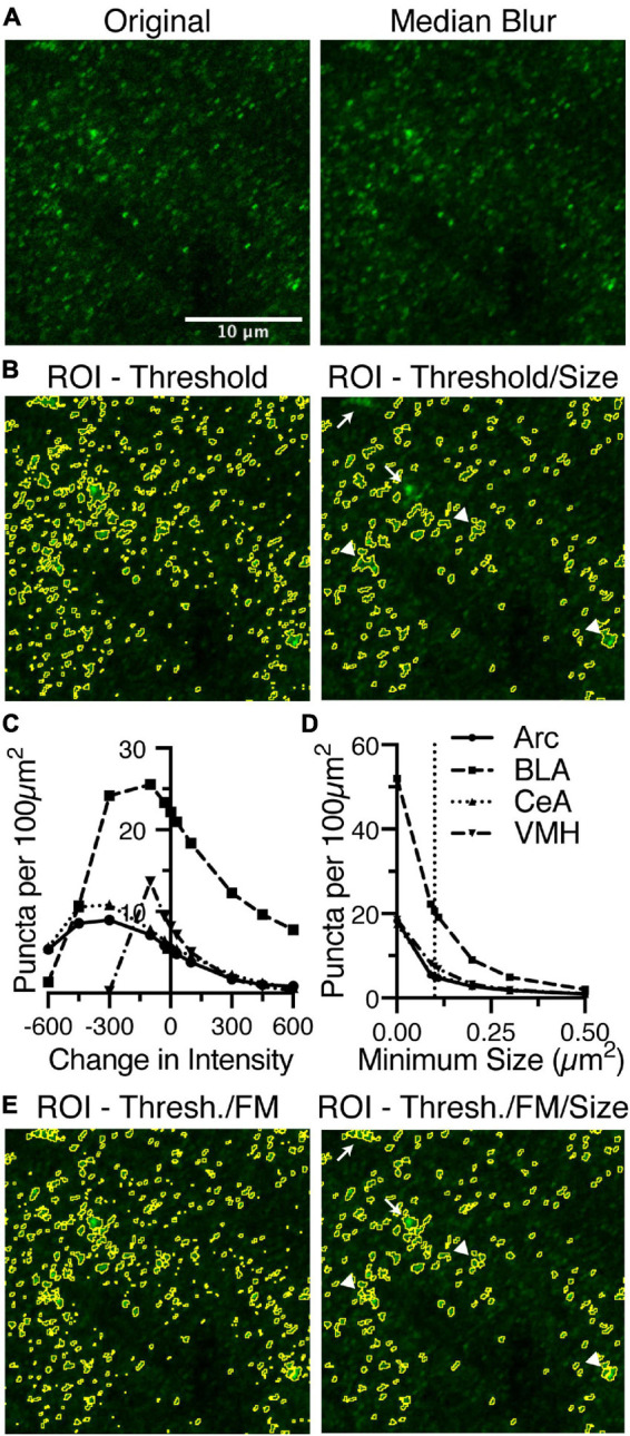
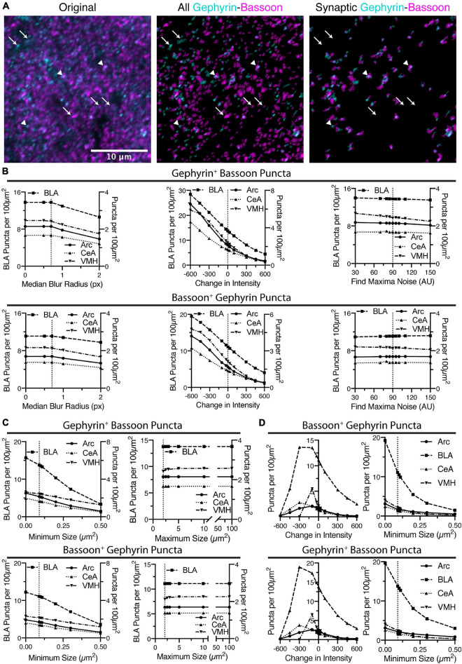
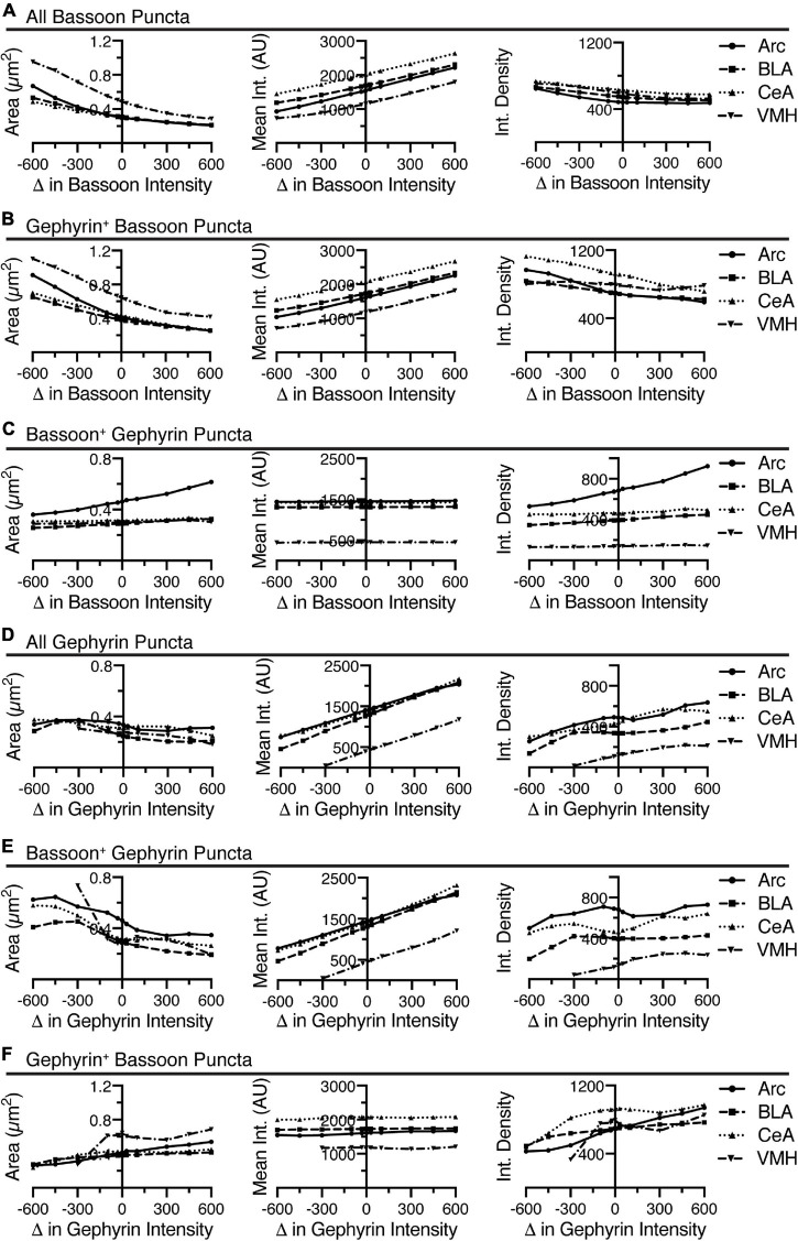
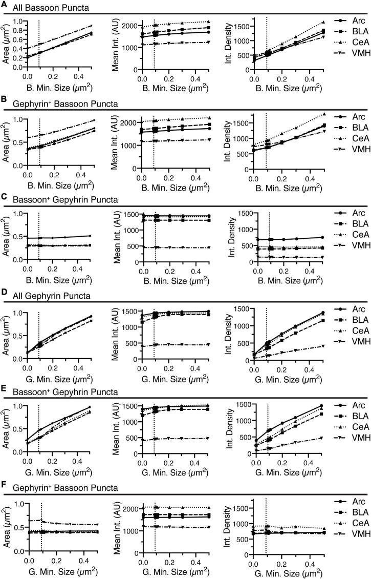
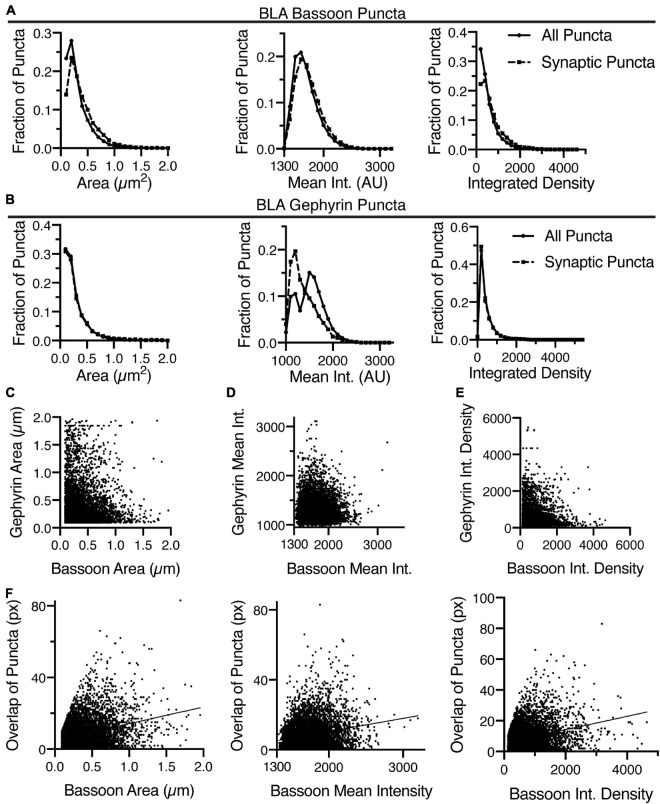
While electron microscopy represents the gold standard for detection of synapses, a number of limitations prevent its broad applicability. A key method for detecting synapses is immunostaining for markers of pre- and post-synaptic proteins, which can infer a synapse based upon the apposition of the two markers. While immunostaining and imaging techniques have improved to allow for identification of synapses in tissue, analysis and identification of these appositions are not facile, and there has been a lack of tools to accurately identify these appositions. Here, we delineate a macro that uses open-source and freely available ImageJ or FIJI for analysis of multichannel, z-stack confocal images. With use of a high magnification with a high NA objective, we outline two methods to identify puncta in either sparsely or densely labeled images. Puncta from each channel are used to eliminate non-apposed puncta and are subsequently linked with their cognate from the other channel. These methods are applied to analysis of a pre-synaptic marker, bassoon, with two different post-synaptic markers, gephyrin and N-methyl-d-aspartate (NMDA) receptor subunit 1 (NR1). Using gephyrin as an inhibitory, post-synaptic scaffolding protein, we identify inhibitory synapses in basolateral amygdala, central amygdala, arcuate and the ventromedial hypothalamus. Systematic variation of the settings identify the parameters most critical for this analysis. Identification of specifically overlapping puncta allows for correlation of morphometry data between each channel. Finally, we extend the analysis to only examine puncta overlapping with a cytoplasmic marker of specific cell types, a distinct advantage beyond electron microscopy. Bassoon puncta are restricted to virally transduced, pedunculopontine tegmental nucleus (PPN) axons expressing yellow fluorescent protein. NR1 puncta are restricted to tyrosine hydroxylase labeled dopaminergic neurons of the substantia nigra pars compacta (SNc). The macro identifies bassoon-NR1 overlap throughout the image, or those only restricted to the PPN-SNc connections. Thus, we have extended the available analysis tools that can be used to study synapses . Our analysis code is freely available and open-source allowing for further innovation.
虽然电子显微镜是检测突触的金标准,但许多限制使其无法广泛应用。检测突触的一种关键方法是免疫染色前突触和后突触蛋白的标志物,根据这两种标志物的接近程度可以推断出突触。虽然免疫染色和成像技术已经得到改进,可以在组织中识别突触,但这些接近点的分析和识别并不容易,也缺乏准确识别这些接近点的工具。在这里,我们描述了一个使用开源和免费的 ImageJ 或 FIJI 分析多通道、z 堆叠共聚焦图像的宏。使用高放大倍率和高数值孔径物镜,我们概述了两种方法来识别稀疏或密集标记图像中的斑点。来自每个通道的斑点用于消除非接近斑点,然后与来自另一个通道的同源斑点连接。这些方法应用于前突触标志物 bassoon 与两个不同的后突触标志物 gephyrin 和 N-甲基-D-天冬氨酸 (NMDA) 受体亚单位 1 (NR1) 的分析。使用 gephyrin 作为抑制性的后突触支架蛋白,我们在基底外侧杏仁核、中央杏仁核、弓状核和腹内侧下丘脑鉴定抑制性突触。系统地改变设置可以确定此分析中最关键的参数。特异性重叠斑点的识别允许在每个通道之间进行形态计量数据的相关性。最后,我们将分析扩展到仅检查与特定细胞类型的细胞质标志物重叠的斑点,这是电子显微镜之外的一个明显优势。Bassoon 斑点仅限于表达黄色荧光蛋白的病毒转导的脚桥核被盖部(PPN)轴突。NR1 斑点仅限于酪氨酸羟化酶标记的黑质致密部(SNc)中的多巴胺能神经元。宏在整个图像中识别 bassoon-NR1 重叠,或者仅局限于 PPN-SNc 连接。因此,我们扩展了可用于研究突触的可用分析工具。我们的分析代码是免费的,开源的,允许进一步的创新。