• 文献检索
  • 文档翻译
  • 深度研究
  • 学术资讯
  • Suppr Zotero 插件Zotero 插件
  • 邀请有礼
  • 套餐&价格
  • 历史记录
应用&插件
Suppr Zotero 插件Zotero 插件浏览器插件Mac 客户端Windows 客户端微信小程序
定价
高级版会员购买积分包购买API积分包
服务
文献检索文档翻译深度研究API 文档MCP 服务
关于我们
关于 Suppr公司介绍联系我们用户协议隐私条款
关注我们

Suppr 超能文献

核心技术专利:CN118964589B侵权必究
粤ICP备2023148730 号-1Suppr @ 2026

文献检索

告别复杂PubMed语法,用中文像聊天一样搜索,搜遍4000万医学文献。AI智能推荐,让科研检索更轻松。

立即免费搜索

文件翻译

保留排版,准确专业,支持PDF/Word/PPT等文件格式,支持 12+语言互译。

免费翻译文档

深度研究

AI帮你快速写综述,25分钟生成高质量综述,智能提取关键信息,辅助科研写作。

立即免费体验

SPECT/CT 评估疑似骨骼病变。

SPECT/CT in the Evaluation of Suspected Skeletal Pathology.

机构信息

Department of Radiology and Imaging Sciences, University of Utah, Salt Lake City, UT 84132, USA.

出版信息

Tomography. 2021 Oct 11;7(4):581-605. doi: 10.3390/tomography7040050.

DOI:10.3390/tomography7040050
PMID:34698290
原文链接:https://pmc.ncbi.nlm.nih.gov/articles/PMC8544734/
Abstract

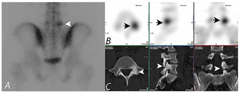
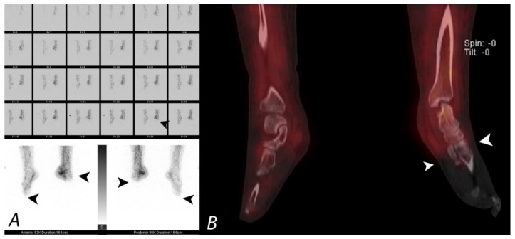
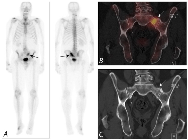
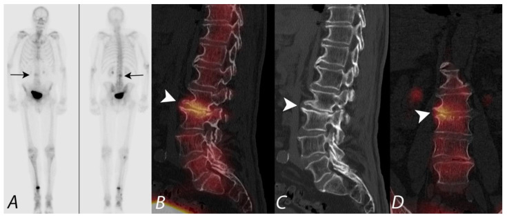
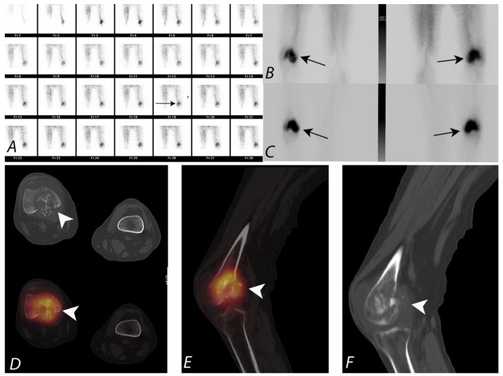
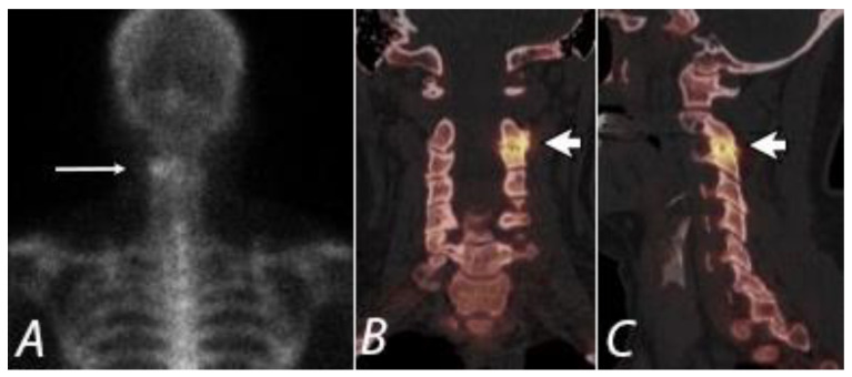
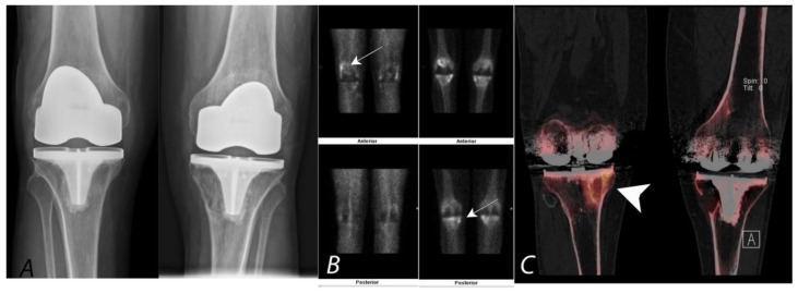
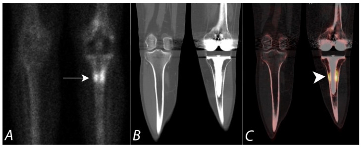
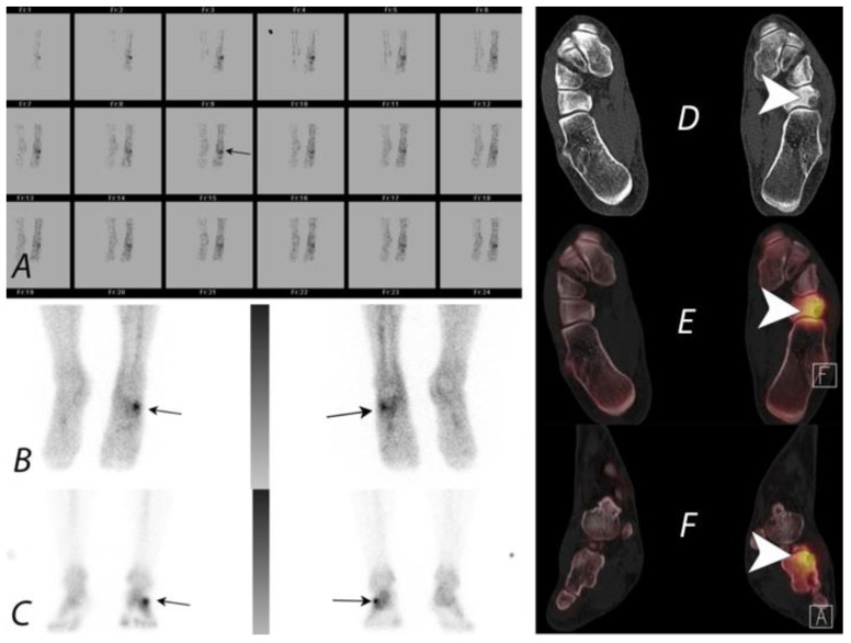
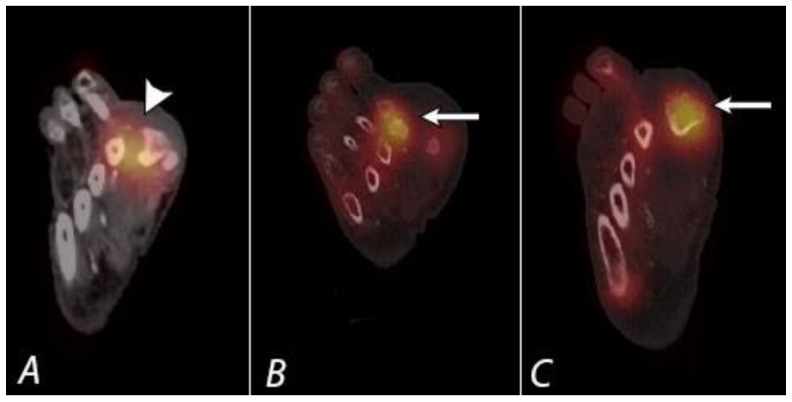
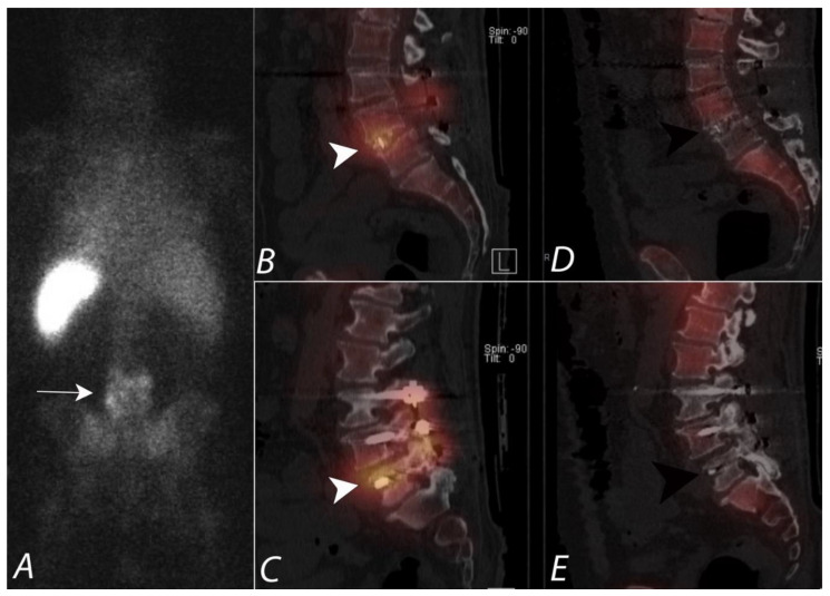
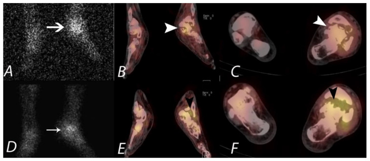
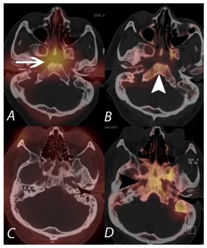
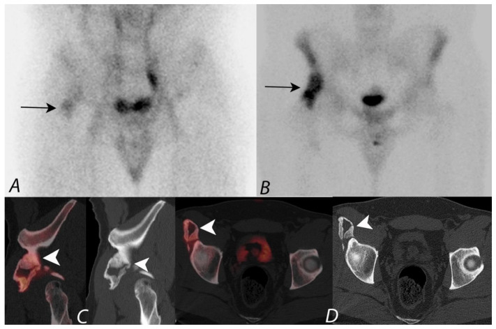
Dedicated multi-slice single-photon emission computed tomography/computed tomography (SPECT/CT) cameras have become widely available and are becoming a mainstay of clinical practice. The integration of SPECT and CT allow for precise anatomic location of scintigraphic findings. Fusion imaging with SPECT/CT can improve both sensitivity and specificity by reducing equivocal interpretation in comparison to planar scintigraphy or SPECT alone. This review article addresses the technique, basic science principles, and applications of integrated SPECT/CT in the evaluation of musculoskeletal pathology.

摘要

专用多层单光子发射计算机断层扫描/计算机断层扫描(SPECT/CT)相机已经广泛应用,并成为临床实践的主要手段。SPECT 和 CT 的整合使得闪烁成像的发现能够精确地定位在解剖位置上。与平面闪烁显像或单独的 SPECT 相比,SPECT/CT 的融合成像可以通过减少不确定的解释来提高敏感性和特异性。本文回顾了 SPECT/CT 在评价肌肉骨骼病变中的技术、基础科学原理和应用。

https://cdn.ncbi.nlm.nih.gov/pmc/blobs/a87a/8544734/ae4405530b37/tomography-07-00050-g017.jpg
https://cdn.ncbi.nlm.nih.gov/pmc/blobs/a87a/8544734/f9ce8e39d729/tomography-07-00050-g001.jpg
https://cdn.ncbi.nlm.nih.gov/pmc/blobs/a87a/8544734/fa8fa3a71d42/tomography-07-00050-g002.jpg
https://cdn.ncbi.nlm.nih.gov/pmc/blobs/a87a/8544734/5c902e751614/tomography-07-00050-g003.jpg
https://cdn.ncbi.nlm.nih.gov/pmc/blobs/a87a/8544734/c9995a406457/tomography-07-00050-g004.jpg
https://cdn.ncbi.nlm.nih.gov/pmc/blobs/a87a/8544734/c543a81ecdf7/tomography-07-00050-g005.jpg
https://cdn.ncbi.nlm.nih.gov/pmc/blobs/a87a/8544734/87697e9377a4/tomography-07-00050-g006.jpg
https://cdn.ncbi.nlm.nih.gov/pmc/blobs/a87a/8544734/89573753b254/tomography-07-00050-g007.jpg
https://cdn.ncbi.nlm.nih.gov/pmc/blobs/a87a/8544734/ad184277eedc/tomography-07-00050-g008.jpg
https://cdn.ncbi.nlm.nih.gov/pmc/blobs/a87a/8544734/80f364cb8cbf/tomography-07-00050-g009.jpg
https://cdn.ncbi.nlm.nih.gov/pmc/blobs/a87a/8544734/5b912cea2c0f/tomography-07-00050-g010.jpg
https://cdn.ncbi.nlm.nih.gov/pmc/blobs/a87a/8544734/279b328ad780/tomography-07-00050-g011.jpg
https://cdn.ncbi.nlm.nih.gov/pmc/blobs/a87a/8544734/8779ddf462a0/tomography-07-00050-g012.jpg
https://cdn.ncbi.nlm.nih.gov/pmc/blobs/a87a/8544734/82020bac02af/tomography-07-00050-g013.jpg
https://cdn.ncbi.nlm.nih.gov/pmc/blobs/a87a/8544734/c26412db9e7d/tomography-07-00050-g014.jpg
https://cdn.ncbi.nlm.nih.gov/pmc/blobs/a87a/8544734/98bb2cfedfbf/tomography-07-00050-g015.jpg
https://cdn.ncbi.nlm.nih.gov/pmc/blobs/a87a/8544734/c63e4d511495/tomography-07-00050-g016.jpg
https://cdn.ncbi.nlm.nih.gov/pmc/blobs/a87a/8544734/ae4405530b37/tomography-07-00050-g017.jpg

相似文献

1
SPECT/CT in the Evaluation of Suspected Skeletal Pathology.SPECT/CT 评估疑似骨骼病变。
Tomography. 2021 Oct 11;7(4):581-605. doi: 10.3390/tomography7040050.
2
SPECT-CT: applications in musculoskeletal radiology.SPECT-CT:在肌肉骨骼放射学中的应用。
Br J Radiol. 2013 Nov;86(1031):20120519. doi: 10.1259/bjr.20120519. Epub 2013 Oct 4.
3
Accuracy of hepatobiliary scintigraphy and added value of SPECT/CT versus planar imaging for diagnosing biliary atresia.肝胆闪烁显像与 SPECT/CT 平面显像诊断胆道闭锁的准确性比较。
Hell J Nucl Med. 2021 May-Aug;24(2):108-113. doi: 10.1967/s002449912350. Epub 2021 Aug 6.
4
Sub 4 minute superfast single-photon emission computed tomography/computed tomography as an add-on for problem-solving in planar bone scintigraphy: a time-saving solution for departments without whole-body single-photon emission computed tomography/computed tomography.4分钟以下超快速单光子发射计算机断层扫描/计算机断层扫描作为平面骨闪烁显像问题解决的附加手段:为没有全身单光子发射计算机断层扫描/计算机断层扫描的科室提供的省时解决方案。
Nucl Med Commun. 2023 May 1;44(5):407-413. doi: 10.1097/MNM.0000000000001677. Epub 2023 Mar 3.
5
Evaluation of the diagnostic value of preoperative sentinel lymph node (SLN) imaging in penile carcinoma patients without palpable inguinal lymph nodes via single photon emission computed tomography/computed tomography (SPECT/CT) as compared to planar scintigraphy.通过单光子发射计算机断层扫描/计算机断层扫描(SPECT/CT)与平面闪烁扫描法比较,评估术前前哨淋巴结(SLN)成像对无可触及腹股沟淋巴结的阴茎癌患者的诊断价值。
Urol Oncol. 2018 Mar;36(3):92.e17-92.e24. doi: 10.1016/j.urolonc.2017.11.012. Epub 2017 Dec 14.
6
Musculoskeletal Spect-Ct: A Pictorial Review.肌肉骨骼系统单光子发射计算机断层扫描-计算机断层扫描:图文综述
J Ayub Med Coll Abbottabad. 2016 Apr-Jun;28(2):427-437.
7
Accuracy of skeletal scintigraphy for the evaluation of mandibular growth disorders: a systematic review.骨骼闪烁显像术评估下颌生长发育障碍的准确性:系统评价。
Int J Oral Maxillofac Surg. 2024 Jun;53(6):482-495. doi: 10.1016/j.ijom.2023.12.006. Epub 2023 Dec 28.
8
Utility of single-photon emission tomography/computed tomography for sentinel lymph node localization in breast cancer patients.单光子发射断层扫描/计算机断层扫描在乳腺癌患者前哨淋巴结定位中的应用价值。
Nucl Med Commun. 2017 Jun;38(6):493-499. doi: 10.1097/MNM.0000000000000676.
9
Single photon emission computed tomography/spiral computed tomography fusion imaging for the diagnosis of bone metastasis in patients with known cancer.单光子发射计算机断层扫描/螺旋计算机断层融合成像在已知癌症患者骨转移诊断中的应用。
Skeletal Radiol. 2010 Feb;39(2):147-53. doi: 10.1007/s00256-009-0764-0.
10
Incremental value of Tc-HYNIC-TOC SPECT/CT over whole-body planar scintigraphy and SPECT in patients with neuroendocrine tumours.锝[Tc] 泮替膦酸盐单光子发射计算机断层显像/计算机断层扫描(Tc-HYNIC-TOC SPECT/CT)相对于全身平面闪烁显像和单光子发射计算机断层扫描(SPECT)在神经内分泌肿瘤患者中的增量价值
Nuklearmedizin. 2017 Jun 12;56(3):97-107. doi: 10.3413/Nukmed-0851-16-10. Epub 2017 Feb 6.

引用本文的文献

1
Outcomes Of Intraarticular Corticosteroid Injections into the Cervical Facet Joint Based on Single-Photon Emission Computed Tomography Imaging.基于单光子发射计算机断层扫描成像的颈椎小关节内注射皮质类固醇的疗效
J Pain Res. 2025 May 14;18:2453-2458. doi: 10.2147/JPR.S522930. eCollection 2025.
2
Prevalence of Degenerative Spinal Hot Spots on Bone Scintigraphy Among Orthopedic Patients.骨科患者中骨闪烁显像上退行性脊柱热点的患病率
Cureus. 2025 Jan 21;17(1):e77779. doi: 10.7759/cureus.77779. eCollection 2025 Jan.
3
Diagnostic criteria for temporomandibular joint osteoarthritis using standardized uptake value in single-photon emission computed tomography-computed tomography.

本文引用的文献

1
Diagnostic Value of Bone SPECT/CT in Patients with Suspected Osteomyelitis.骨SPECT/CT在疑似骨髓炎患者中的诊断价值
Mol Imaging Radionucl Ther. 2019 Sep 6;28(3):89-95. doi: 10.4274/mirt.galenos.2019.20053.
2
Clinical relevance of persistent postoperative pain after total hip replacement - a prospective observational cohort study.全髋关节置换术后持续性疼痛的临床相关性——一项前瞻性观察队列研究。
J Pain Res. 2017 Sep 7;10:2183-2193. doi: 10.2147/JPR.S137892. eCollection 2017.
3
Bone SPECT/CT in the postoperative spine: a focus on spinal fusion.
使用单光子发射计算机断层扫描-计算机断层扫描中的标准化摄取值诊断颞下颌关节骨关节炎的标准
Sci Rep. 2024 Dec 30;14(1):31569. doi: 10.1038/s41598-024-71639-1.
4
Short-Term Outcomes of Intraarticular Corticosteroid Injection into the Lumbar Facet Joint According to the Findings of Single-Photon Emission Computed Tomography Imaging.根据单光子发射计算机断层扫描成像结果进行腰椎小关节腔内注射皮质类固醇的短期疗效
J Pain Res. 2024 Jul 9;17:2357-2362. doi: 10.2147/JPR.S467549. eCollection 2024.
5
Coronal Deformity is Associated With Uptake on Single Photon Emission Computed Tomography in Patients With Low Back Pain.在腰痛患者中,冠状面畸形与单光子发射计算机断层扫描中的摄取有关。
Global Spine J. 2025 Apr;15(3):1783-1791. doi: 10.1177/21925682241265302. Epub 2024 Jun 19.
6
Verification of image quality improvement of low-count bone scintigraphy using deep learning.使用深度学习验证低计数骨闪烁显像的图像质量改善情况。
Radiol Phys Technol. 2024 Mar;17(1):269-279. doi: 10.1007/s12194-023-00776-5. Epub 2024 Feb 10.
7
Radiological Insights into Sacroiliitis: A Narrative Review.骶髂关节炎的影像学见解:一篇叙述性综述
Clin Pract. 2024 Jan 3;14(1):106-121. doi: 10.3390/clinpract14010009.
8
INHBA gene silencing inhibits proliferation, migration, and invasion of osteosarcoma cells by repressing TGF-β signaling pathway activation.INHBA 基因沉默通过抑制 TGF-β 信号通路的激活抑制骨肉瘤细胞的增殖、迁移和侵袭。
J Orthop Surg Res. 2023 Nov 8;18(1):848. doi: 10.1186/s13018-023-04330-2.
9
A Review on the Usage of Bone Single-Photon Emission Computed Tomography/Computed Tomography in Detecting Skeletal Metastases in the Post-COVID-19 Era: Is it Time to Ditch Planar and Single-Photon Emission Computed Tomography only Gamma Camera Systems?关于骨单光子发射计算机断层扫描/计算机断层扫描在新冠疫情后时代检测骨转移中的应用综述:是时候摒弃仅使用平面和单光子发射计算机断层扫描的伽马相机系统了吗?
Indian J Nucl Med. 2023 Apr-Jun;38(2):191-200. doi: 10.4103/ijnm.ijnm_142_22. Epub 2023 Jun 8.
10
Evaluation of optimal single-photon emission computed tomography reference value and three-dimensional mandibular growth pattern in 54 Chinese unilateral condylar hyperplasia patients.54 例中国单侧髁突肥大患者最佳单光子发射计算机断层扫描参考值与下颌骨三维生长模式的评估。
Head Face Med. 2023 May 19;19(1):18. doi: 10.1186/s13005-023-00365-2.
骨 SPECT/CT 在脊柱术后:关注脊柱融合。
Eur J Nucl Med Mol Imaging. 2017 Nov;44(12):2094-2104. doi: 10.1007/s00259-017-3765-6. Epub 2017 Jul 5.
4
SPECT/CT: an update on technological developments and clinical applications.单光子发射计算机断层扫描/计算机断层扫描:技术发展与临床应用的最新进展
Br J Radiol. 2018 Jan;91(1081):20160402. doi: 10.1259/bjr.20160402. Epub 2017 Jan 16.
5
Atypical Fractures Following Bisphosphonate Therapy.双膦酸盐治疗后的非典型骨折
Semin Musculoskelet Radiol. 2016 Sep;20(4):376-381. doi: 10.1055/s-0036-1593766. Epub 2016 Nov 14.
6
Frostbite: Spectrum of Imaging Findings and Guidelines for Management.冻伤:影像学表现谱及处理指南
Radiographics. 2016 Nov-Dec;36(7):2154-2169. doi: 10.1148/rg.2016160045. Epub 2016 Aug 5.
7
Total Hip Arthroplasty in 6690 Patients with Inflammatory Arthritis: Effect of Medical Comorbidities and Age on Early Mortality.6690例炎性关节炎患者的全髋关节置换术:内科合并症和年龄对早期死亡率的影响
J Rheumatol. 2016 Jul;43(7):1320-7. doi: 10.3899/jrheum.151287. Epub 2016 May 1.
8
Accuracy of bone SPECT/CT for identifying hardware loosening in patients who underwent lumbar fusion with pedicle screws.骨SPECT/CT在接受椎弓根螺钉腰椎融合术患者中识别内固定松动的准确性。
Eur J Nucl Med Mol Imaging. 2016 Feb;43(2):349-354. doi: 10.1007/s00259-015-3158-7. Epub 2015 Aug 13.
9
Clinical value of SPECT/CT in the painful total knee arthroplasty (TKA): a prospective study in a consecutive series of 100 TKA.SPECT/CT在疼痛性全膝关节置换术(TKA)中的临床价值:对连续100例TKA患者的前瞻性研究
Eur J Nucl Med Mol Imaging. 2015 Nov;42(12):1869-82. doi: 10.1007/s00259-015-3095-5. Epub 2015 Jun 6.
10
Hybrid SPECT/CT for the assessment of a painful hip after uncemented total hip arthroplasty.混合单光子发射计算机断层扫描/计算机断层扫描用于评估非骨水泥型全髋关节置换术后疼痛性髋关节。
BMC Med Imaging. 2015 Jun 2;15:18. doi: 10.1186/s12880-015-0056-1.