Karolinska Institutet Karolinska University Hospital, Stockholm, Sweden.
Royal London Hospital, Barts Healthcare NHS Trust, London, UK.
Sci Rep. 2021 Oct 26;11(1):21123. doi: 10.1038/s41598-021-00532-y.
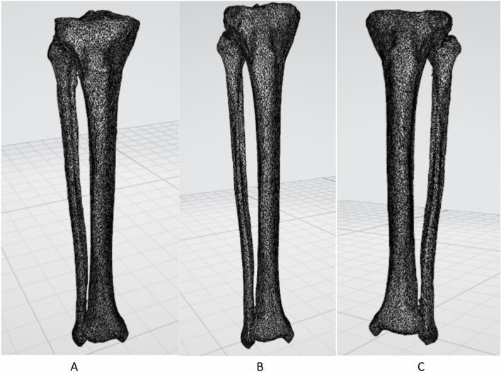
Rotational deformities following intramedullary (IM) nailing of tibia has a reported incidence of as high as 20%. Common techniques to measure deformities following IM nailing of tibia are either based on clinical assessment, plain X-rays or Computed Tomography (CT) comparing the treated leg with the uninjured contralateral side. All these techniques are based on examiners manual calculation inherently subject to bias. Following our previous rigorous motion analysis and symmetry studies on hemi pelvises, femurs and orthopaedic implants, we aimed to introduce a novel fully digital technique to measure rotational deformities in the lower legs. Following formal institutional approval from the Imperial College, CT images of 10 pairs of human lower legs were retrieved. Images were anonymized and uploaded to a research server. Three dimensional CT images of the lower legs were bilaterally reconstructed. CT-based motion analysis (CTMA) was used and the mirrored images of the left side were merged with the right side proximally as stationary and distally as moving objects. Discrepancies in translation and rotation were automatically calculated. Our study population had a mean age of 54 ± 20 years. There were six males and four females. We observed a greater variation in translation (mm) of Centre of Mass (COM) in sagittal plane (95% CI - 2.959-.292) which was also presented as rotational difference alongside the antero-posterior direction or Y axis (95% CI .370-1.035). In other word the right lower legs in our study were more likely to be in varus compared to the left side. However, there were no statistically significant differences in coronal or axial planes. Using our proposed fully digital technique we found that lower legs of the human adults were symmetrical in axial and coronal plane. We found sagittal plane differences which need further addressing in future using bigger sample size. Our novel recommended technique is fully digital and commercially available. This new technique can be useful in clinical practice addressing rotational deformities following orthopaedic surgical intervention. This new technique can substitute the previously introduced techniques.
胫骨髓内钉固定后发生旋转畸形的发生率高达 20%。目前常用的测量胫骨髓内钉固定后畸形的方法有临床评估、普通 X 线片或与未受伤对侧比较的计算机断层扫描(CT)。所有这些技术都是基于检查者的手动计算,固有偏差。在我们之前对骨盆半部分、股骨和骨科植入物进行了严格的运动分析和对称性研究之后,我们旨在引入一种新的完全数字化技术来测量小腿的旋转畸形。在得到帝国理工学院的正式机构批准后,我们检索了 10 对人类小腿的 CT 图像。对图像进行了匿名处理,并上传到研究服务器。双侧重建小腿的三维 CT 图像。使用基于 CT 的运动分析(CTMA),将左侧镜像图像与右侧近端作为固定对象,远端作为移动对象进行合并。自动计算平移和旋转的差异。我们的研究人群平均年龄为 54±20 岁。有 6 名男性和 4 名女性。我们观察到矢状面质心(COM)的平移(mm)差异较大(95%CI -2.959-.292),同时也表现为前-后方向或 Y 轴的旋转差异(95%CI.370-1.035)。换句话说,与左侧相比,我们研究中的右侧小腿更有可能出现内翻。然而,冠状面或轴面没有统计学差异。使用我们提出的完全数字化技术,我们发现成年人的小腿在冠状面和矢状面是对称的。我们发现了矢状面的差异,需要在未来使用更大的样本量进一步研究。我们新推荐的技术是完全数字化的,并且是商业可用的。这种新技术在处理骨科手术后的旋转畸形方面在临床实践中可能很有用。这种新技术可以替代以前介绍的技术。