Cha Misun, Jin Yuan-Zhe, Park Jin Wook, Lee Kyung Mee, Han Shi Huan, Choi Byung Sun, Lee Jae Hyup
Biotechnology Institute, Medifab Co. LTD., 70, Dusan-ro, Doksan-dong, Geumcheon-gu, Seoul, 085-84, South Korea.
Department of Orthopedic Surgery, SMG-SNU Boramae Medical Center, 39 Boramae Gil, Dongjak-Gu, Seoul, 156-707, South Korea.
Biomater Res. 2021 Oct 27;25(1):35. doi: 10.1186/s40824-021-00233-7.
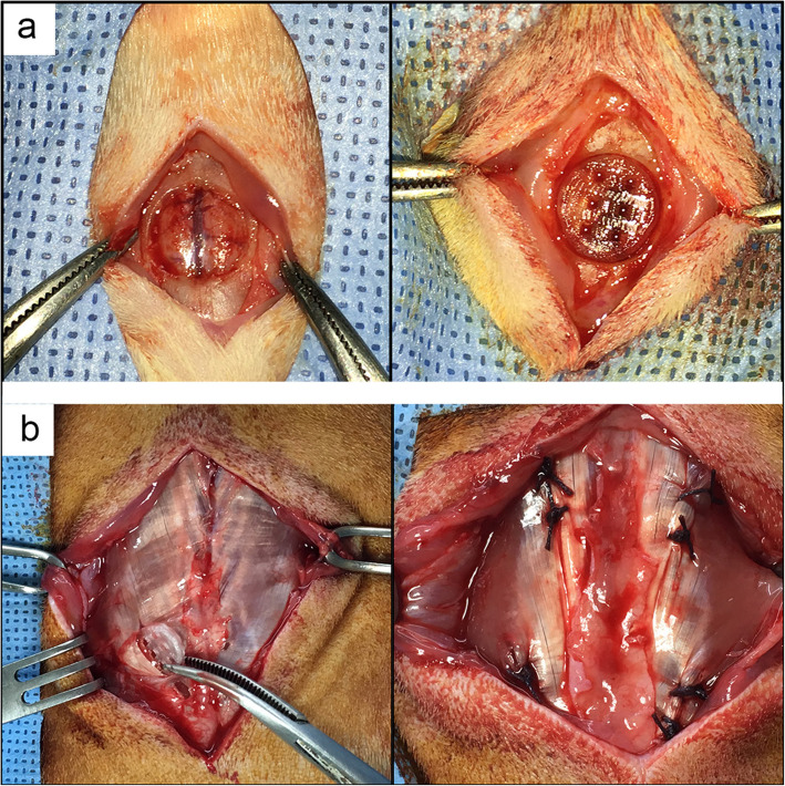
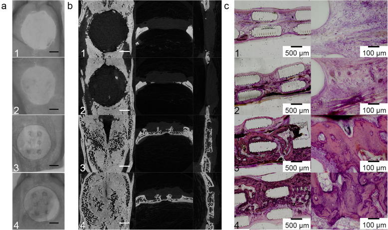
Critical bone defects remain challenges for clinicians, which cannot heal spontaneously and require medical intervention. Following the development of three-dimensional (3D) printing technology is widely used in bone tissue engineering for its outstanding customizability. The 3D printed scaffolds were usually accompanied with growth factors, such as bone morphometric protein 2 (BMP-2), whose effects have been widely investigated on bone regeneration. We previously fabricated and investigated the effect of a polylactic acid (PLA) cage/Biogel scaffold as a carrier of BMP-2. In this study, we furtherly investigated the effect of another shape of PLA cage/Biogel scaffold as a carrier of BMP-2 in a rat calvaria defect model and an ectopic ossification (EO) model.
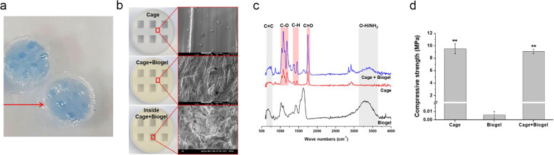
The PLA scaffold was printed with a basic commercial 3D printer, and the PLA scaffold was combined with gelatin and alginate-based Biogel and BMP-2 to induce bone regeneration. The experimental groups were divided into PLA scaffold, PLA scaffold with Biogel, PLA scaffold filled with BMP-2, and PLA scaffold with Biogel and BMP-2 and were tested both in vitro and in vivo. One-way ANOVA with Bonferroni post-hoc analysis was used to determine whether statistically significant difference exists between groups.
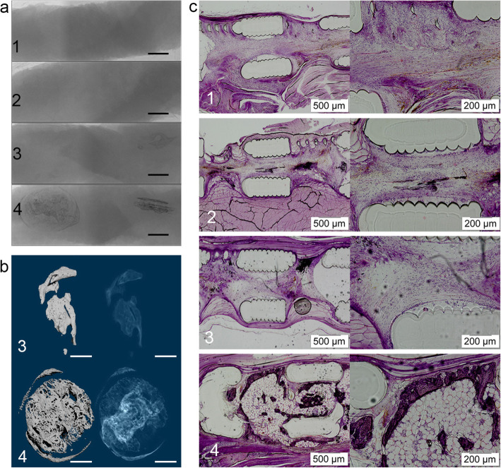
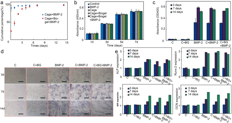
The in vitro results showed the cage/Biogel scaffold released BMP-2 with an initial burst release and followed by a sustained slow-release pattern. The released BMP-2 maintained its osteoinductivity for at least 14 days. The in vivo results showed the cage/Biogel/BMP-2 group had the highest bone regeneration in the rat calvarial defect model and EO model. Especially, the bone regenerated more regularly in the EO model at the implanted sites, which indicated the cage/Biogel had an outstanding ability to control the shape of regenerated bone.
In conclusion, the 3D printed PLA cage/Biogel scaffold system was proved to be a proper carrier for BMP-2 that induced significant bone regeneration and induced bone formation following the designed shape.
严重骨缺损仍是临床医生面临的挑战,这些骨缺损无法自发愈合,需要医学干预。随着三维(3D)打印技术的发展,因其出色的可定制性而被广泛应用于骨组织工程。3D打印支架通常会结合生长因子,如骨形态发生蛋白2(BMP-2),其对骨再生的作用已得到广泛研究。我们之前制备并研究了聚乳酸(PLA)笼/生物凝胶支架作为BMP-2载体的效果。在本研究中,我们进一步研究了另一种形状的PLA笼/生物凝胶支架作为BMP-2载体在大鼠颅骨缺损模型和异位骨化(EO)模型中的效果。
使用基本的商用3D打印机打印PLA支架,并将PLA支架与基于明胶和藻酸盐的生物凝胶以及BMP-2结合以诱导骨再生。实验组分为PLA支架组、带生物凝胶的PLA支架组、填充BMP-2的PLA支架组以及带生物凝胶和BMP-2的PLA支架组,并在体外和体内进行测试。采用单因素方差分析和Bonferroni事后分析来确定组间是否存在统计学上的显著差异。
体外结果显示,笼状/生物凝胶支架以初始爆发释放然后持续缓慢释放的模式释放BMP-2。释放的BMP-2至少在14天内保持其骨诱导活性。体内结果显示,在大鼠颅骨缺损模型和EO模型中,笼状/生物凝胶/BMP-2组的骨再生效果最佳。特别是,在EO模型的植入部位,再生骨的形态更规则,这表明笼状/生物凝胶具有出色的控制再生骨形状的能力。
总之,3D打印的PLA笼/生物凝胶支架系统被证明是BMP-2的合适载体,可诱导显著的骨再生并按照设计形状诱导骨形成。