Department of Pharmacy, School of Applied Sciences, University of Huddersfield, Queensgate, Huddersfield, HD1 3DH, UK.
Mol Neurobiol. 2022 Jan;59(1):445-458. doi: 10.1007/s12035-021-02593-6. Epub 2021 Oct 28.
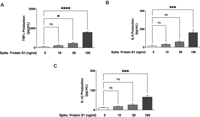
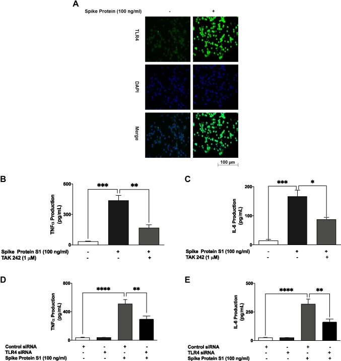
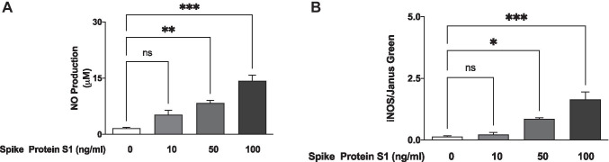
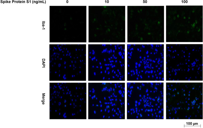
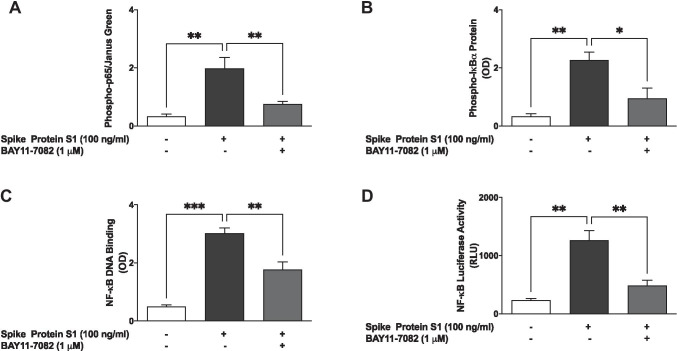
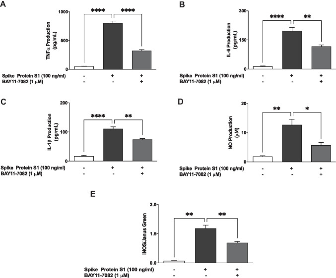
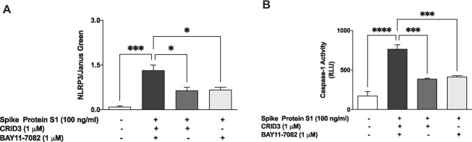
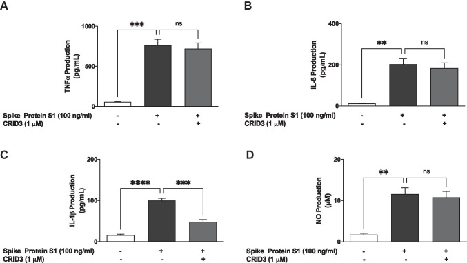
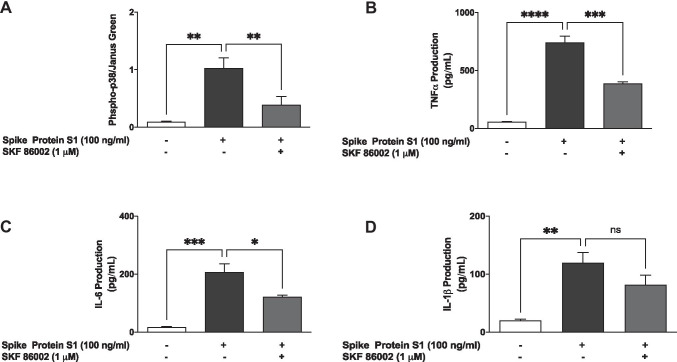
In addition to respiratory complications produced by SARS-CoV-2, accumulating evidence suggests that some neurological symptoms are associated with the disease caused by this coronavirus. In this study, we investigated the effects of the SARS-CoV-2 spike protein S1 stimulation on neuroinflammation in BV-2 microglia. Analyses of culture supernatants revealed an increase in the production of TNF-α, IL-6, IL-1β and iNOS/NO. S1 also increased protein levels of phospho-p65 and phospho-IκBα, as well as enhanced DNA binding and transcriptional activity of NF-κB. These effects of the protein were blocked in the presence of BAY11-7082 (1 µM). Exposure of S1 to BV-2 microglia also increased the protein levels of NLRP3 inflammasome and enhanced caspase-1 activity. Increased protein levels of p38 MAPK was observed in BV-2 microglia stimulated with the spike protein S1 (100 ng/ml), an action that was reduced in the presence of SKF 86,002 (1 µM). Results of immunofluorescence microscopy showed an increase in TLR4 protein expression in S1-stimulated BV-2 microglia. Furthermore, pharmacological inhibition with TAK 242 (1 µM) and transfection with TLR4 small interfering RNA resulted in significant reduction in TNF-α and IL-6 production in S1-stimulated BV-2 microglia. These results have provided the first evidence demonstrating S1-induced neuroinflammation in BV-2 microglia. We propose that induction of neuroinflammation by this protein in the microglia is mediated through activation of NF-κB and p38 MAPK, possibly as a result of TLR4 activation. These results contribute to our understanding of some of the mechanisms involved in CNS pathologies of SARS-CoV-2.
除了由 SARS-CoV-2 引起的呼吸并发症外,越来越多的证据表明,这种冠状病毒引起的疾病与一些神经症状有关。在这项研究中,我们研究了 SARS-CoV-2 刺突蛋白 S1 刺激对 BV-2 小胶质细胞神经炎症的影响。对培养上清液的分析显示,TNF-α、IL-6、IL-1β 和 iNOS/NO 的产生增加。S1 还增加了磷酸化 p65 和磷酸化 IκBα 的蛋白水平,并增强了 NF-κB 的 DNA 结合和转录活性。这些蛋白的作用在存在 BAY11-7082(1 μM)时被阻断。S1 暴露于 BV-2 小胶质细胞也增加了 NLRP3 炎症小体的蛋白水平,并增强了 caspase-1 活性。在受刺突蛋白 S1(100ng/ml)刺激的 BV-2 小胶质细胞中观察到 p38 MAPK 蛋白水平升高,而在存在 SKF 86,002(1 μM)时,该作用降低。免疫荧光显微镜的结果显示,在 S1 刺激的 BV-2 小胶质细胞中 TLR4 蛋白表达增加。此外,用 TAK 242(1 μM)进行药理学抑制和用 TLR4 小干扰 RNA 转染导致 S1 刺激的 BV-2 小胶质细胞中 TNF-α 和 IL-6 产生的显著减少。这些结果首次提供了证据,证明 S1 诱导了 BV-2 小胶质细胞中的神经炎症。我们提出,这种蛋白在小胶质细胞中诱导的神经炎症是通过 NF-κB 和 p38 MAPK 的激活介导的,可能是由于 TLR4 的激活。这些结果有助于我们理解 SARS-CoV-2 引起的中枢神经系统病变的一些机制。