School of Medical Information & Engineering, Xuzhou Medical University, Xuzhou 221004, China.
Department of Information Center, Weihai Ocean Vocational College, Rongcheng 264300, China.
J Healthc Eng. 2021 Oct 27;2021:8769652. doi: 10.1155/2021/8769652. eCollection 2021.
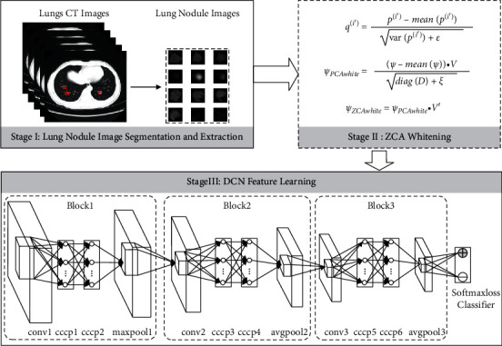
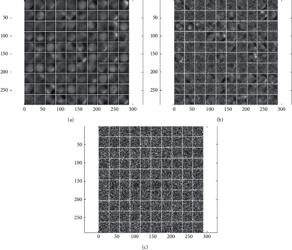
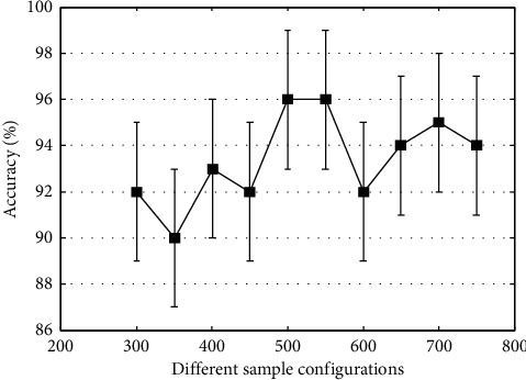
With the rapid development of detection technology, CT imaging technology has been widely used in the early clinical diagnosis of lung nodules. However, accurate assessment of the nature of the nodule remains a challenging task due to the subjective nature of the radiologist. With the increasing amount of publicly available lung image data, it has become possible to use convolutional neural networks for benign and malignant classification of lung nodules. However, as the network depth increases, network training methods based on gradient descent usually lead to gradient dispersion. Therefore, we propose a novel deep convolutional network approach to classify the benignity and malignancy of lung nodules. Firstly, we segmented, extracted, and performed zero-phase component analysis whitening on images of lung nodules. Then, a multilayer perceptron was introduced into the structure to construct a deep convolutional network. Finally, the minibatch stochastic gradient descent method with a momentum coefficient is used to fine-tune the deep convolutional network to avoid the gradient dispersion. The 750 lung nodules in the lung image database are used for experimental verification. Classification accuracy of the proposed method can reach 96.0%. The experimental results show that the proposed method can provide an objective and efficient aid to solve the problem of classifying benign and malignant lung nodules in medical images.
随着检测技术的飞速发展,CT 成像技术已广泛应用于肺结节的早期临床诊断。然而,由于放射科医生的主观性,准确评估结节的性质仍然是一项具有挑战性的任务。随着越来越多的公共肺部图像数据的出现,已经可以使用卷积神经网络对肺结节进行良性和恶性分类。然而,随着网络深度的增加,基于梯度下降的网络训练方法通常会导致梯度弥散。因此,我们提出了一种新的深度卷积网络方法来对肺结节的良恶性进行分类。首先,我们对肺结节的图像进行分割、提取和零相位分量分析白化。然后,引入多层感知器到结构中,构建一个深度卷积网络。最后,使用带有动量系数的小批量随机梯度下降法来微调深度卷积网络,以避免梯度弥散。在肺图像数据库中的 750 个肺结节用于实验验证。所提出方法的分类精度可达到 96.0%。实验结果表明,该方法可以为解决医学图像中良性和恶性肺结节的分类问题提供客观、高效的辅助。