Texas A&M College of Dentistry Center for Craniofacial Research and Diagnosis, United States.
Texas A&M College of Dentistry Center for Craniofacial Research and Diagnosis, United States.
J Struct Biol. 2021 Dec;213(4):107809. doi: 10.1016/j.jsb.2021.107809. Epub 2021 Nov 6.
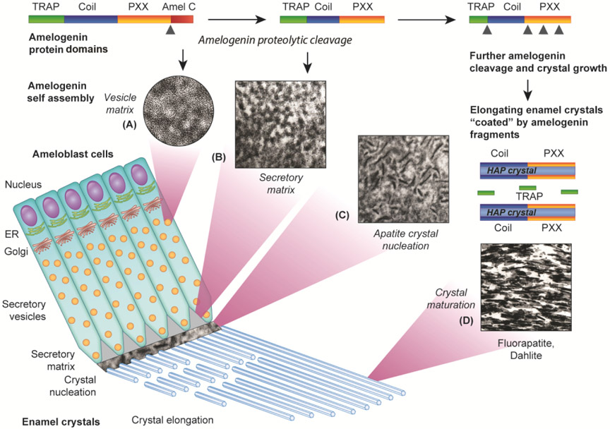
During enamel formation, the organic enamel protein matrix interacts with calcium phosphate minerals to form elongated, parallel, and bundled enamel apatite crystals of extraordinary hardness and biomechanical resilience. The enamel protein matrix consists of unique enamel proteins such as amelogenin, ameloblastin, and enamelin, which are secreted by highly specialized cells called ameloblasts. The ameloblasts also facilitate calcium and phosphate ion transport toward the enamel layer. Within ameloblasts, enamel proteins are transported as a polygonal matrix with 5 nm subunits in secretory vesicles. Upon expulsion from the ameloblasts, the enamel protein matrix is re-organized into 20 nm subunit compartments. Enamel matrix subunit compartment assembly and expansion coincide with C-terminal cleavage by the MMP20 enamel protease and N-terminal amelogenin self-assembly. Upon enamel crystal precipitation, the enamel protein phase is reconfigured to surround the elongating enamel crystals and facilitate their elongation in C-axis direction. At this stage of development, and upon further amelogenin cleavage, central and polyproline-rich fragments of the amelogenin molecule associate with the growing mineral crystals through a process termed "shedding", while hexagonal apatite crystals fuse in longitudinal direction. Enamel protein sheath-coated enamel "dahlite" crystals continue to elongate until a dense bundle of parallel apatite crystals is formed, while the enamel matrix is continuously degraded by proteolytic enzymes. Together, these insights portrait enamel mineral nucleation and growth as a complex and dynamic set of interactions between enamel proteins and mineral ions that facilitate regularly seeded apatite growth and parallel enamel crystal elongation.
在釉质形成过程中,有机釉质蛋白基质与磷酸钙矿物质相互作用,形成具有非凡硬度和生物力学弹性的细长、平行和束状釉质磷灰石晶体。釉质蛋白基质由独特的釉质蛋白组成,如釉原蛋白、釉蛋白和釉质蛋白,这些蛋白由高度特化的成釉细胞分泌。成釉细胞还促进钙和磷酸盐离子向釉质层的运输。在成釉细胞内,釉质蛋白作为一个具有 5nm 亚基的多角形基质在分泌小泡中运输。从成釉细胞排出后,釉质蛋白基质重新组织成 20nm 亚基隔室。釉质基质亚基隔室的组装和扩展与 MMP20 釉质蛋白酶的 C 末端切割和 N 末端釉原蛋白自组装同时发生。在釉质晶体沉淀后,釉质蛋白相被重新配置以包围生长的釉质晶体,并促进它们在 C 轴方向上的伸长。在这个发育阶段,以及进一步的釉原蛋白切割后,釉原蛋白分子的中心和富含脯氨酸的片段通过称为“脱落”的过程与生长的矿物晶体结合,而六方磷灰石晶体在纵向融合。釉质蛋白鞘包裹的釉质“dahlite”晶体继续伸长,直到形成致密的平行磷灰石晶体束,而釉质基质不断被蛋白酶降解。这些见解共同描绘了釉质矿物成核和生长是一个复杂而动态的过程,涉及釉质蛋白和矿物离子之间的相互作用,促进了有序的磷灰石生长和平行釉质晶体伸长。