Department of Neurosurgery, University Hospital, Rennes, France.
INSERM U1242, University of Rennes, Rennes, France.
J Cell Mol Med. 2021 Dec;25(23):10846-10856. doi: 10.1111/jcmm.16902. Epub 2021 Nov 12.
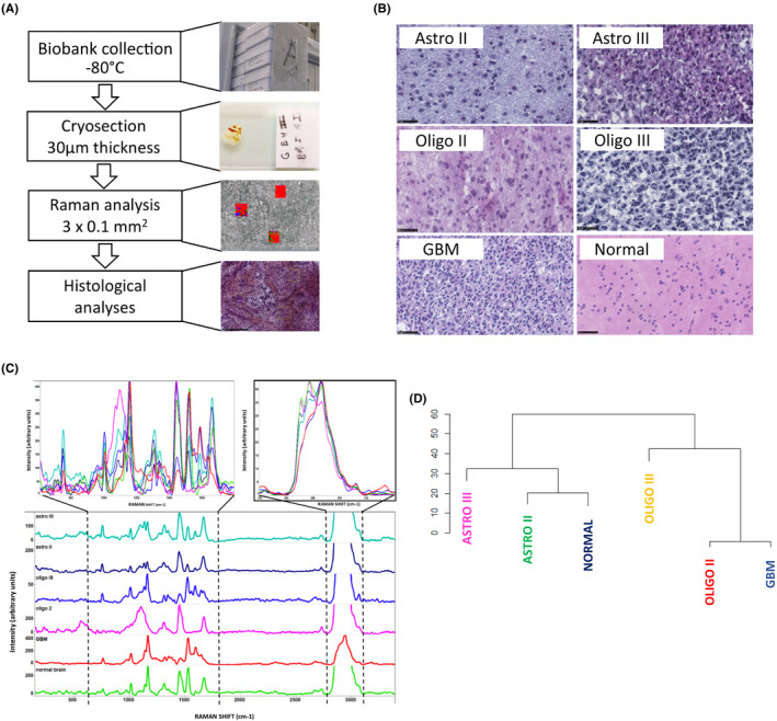
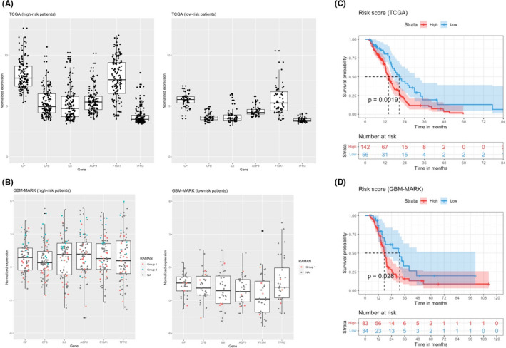
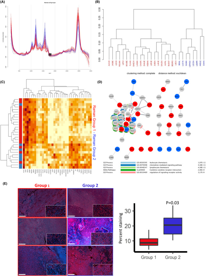
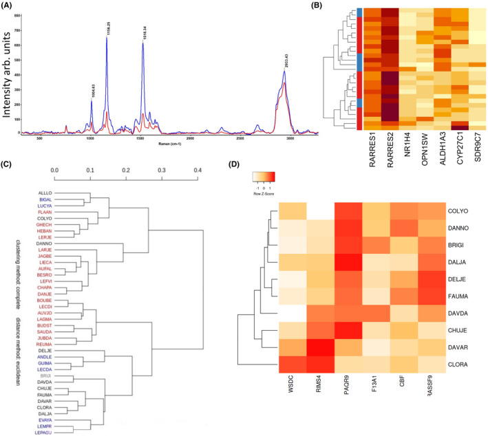
Raman spectroscopy is an imaging technique that has been applied to assess molecular compositions of living cells to characterize cell types and states. However, owing to the diverse molecular species in cells and challenges of assigning peaks to specific molecules, it has not been clear how to interpret cellular Raman spectra. Here, we provide firm evidence that cellular Raman spectra (RS) and transcriptomic profiles of glioblastoma can be computationally connected and thus interpreted. We find that the dimensions of high-dimensional RS and transcriptomes can be reduced and connected linearly through a shared low-dimensional subspace. Accordingly, we were able to predict global gene expression profiles by applying the calculated transformation matrix to Raman spectra and vice versa. From these analyses, we extract a minimal gene expression signature associated with specific RS profiles and predictive of disease outcome.
拉曼光谱是一种成像技术,已被应用于评估活细胞的分子组成,以鉴定细胞类型和状态。然而,由于细胞中存在多种分子物种,以及为特定分子分配峰的困难,因此尚不清楚如何解释细胞拉曼光谱。在这里,我们提供了确凿的证据,表明可以通过计算将脑胶质瘤的细胞拉曼光谱(RS)和转录组图谱联系起来,并进行解释。我们发现,高维 RS 和转录组的维度可以通过共享的低维子空间进行线性缩减和连接。因此,我们可以通过将计算出的变换矩阵应用于拉曼光谱来预测全局基因表达谱,反之亦然。通过这些分析,我们提取出与特定 RS 谱相关并可预测疾病结果的最小基因表达特征。