Riva Marco, Sciortino Tommaso, Secoli Riccardo, D'Amico Ester, Moccia Sara, Fernandes Bethania, Conti Nibali Marco, Gay Lorenzo, Rossi Marco, De Momi Elena, Bello Lorenzo
Department of Medical Biotechnology and Translational Medicine, Università Degli Studi di Milano, 20122 Milan, Italy.
Unit of Oncological Neurosurgery, Humanitas Clinical and Research Center-IRCCS, 20089 Rozzano, Italy.
Cancers (Basel). 2021 Mar 3;13(5):1073. doi: 10.3390/cancers13051073.
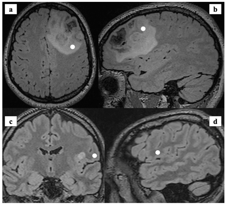
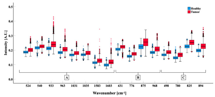
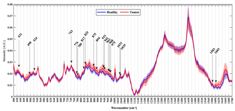
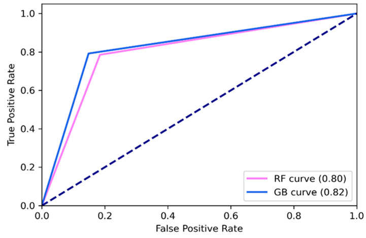
Identifying tumor cells infiltrating normal-appearing brain tissue is critical to achieve a total glioma resection. Raman spectroscopy (RS) is an optical technique with potential for real-time glioma detection. Most RS reports are based on formalin-fixed or frozen samples, with only a few studies deployed on fresh untreated tissue. We aimed to probe RS on untreated brain biopsies exploring novel Raman bands useful in distinguishing glioma and normal brain tissue. Sixty-three fresh tissue biopsies were analyzed within few minutes after resection. A total of 3450 spectra were collected, with 1377 labelled as Healthy and 2073 as Tumor. Machine learning methods were used to classify spectra compared to the histo-pathological standard. The algorithms extracted information from 60 different Raman peaks identified as the most representative among 135 peaks screened. We were able to distinguish between tumor and healthy brain tissue with accuracy and precision of 83% and 82%, respectively. We identified 19 new Raman shifts with known biological significance. Raman spectroscopy was effective and accurate in discriminating glioma tissue from healthy brain ex-vivo in fresh samples. This study added new spectroscopic data that can contribute to further develop Raman Spectroscopy as an intraoperative tool for in-vivo glioma detection.
识别浸润正常脑组织的肿瘤细胞对于实现胶质瘤全切至关重要。拉曼光谱(RS)是一种具有实时检测胶质瘤潜力的光学技术。大多数RS报告基于福尔马林固定或冷冻样本,仅有少数研究应用于新鲜未处理组织。我们旨在对未经处理的脑活检组织进行RS检测,探索有助于区分胶质瘤和正常脑组织的新型拉曼谱带。63份新鲜组织活检样本在切除后几分钟内进行了分析。共收集了3450个光谱,其中1377个标记为健康组织,2073个标记为肿瘤组织。与组织病理学标准相比,使用机器学习方法对光谱进行分类。算法从筛选出的135个峰中确定的60个最具代表性的不同拉曼峰中提取信息。我们能够分别以83%的准确率和82%的精确率区分肿瘤组织和健康脑组织。我们识别出19个具有已知生物学意义的新拉曼位移。拉曼光谱在新鲜样本中离体区分胶质瘤组织和健康脑组织方面有效且准确。本研究增加了新的光谱数据,有助于进一步将拉曼光谱开发为术中体内胶质瘤检测工具。