Panman Jessica L, To Yang Yang, van der Ende Emma L, Poos Jackie M, Jiskoot Lize C, Meeter Lieke H H, Dopper Elise G P, Bouts Mark J R J, van Osch Matthias J P, Rombouts Serge A R B, van Swieten John C, van der Grond Jeroen, Papma Janne M, Hafkemeijer Anne
Department of Radiology, Leiden University Medical Center, Leiden, Netherlands.
Department of Neurology, Erasmus University Medical Center Rotterdam, Rotterdam, Netherlands.
Front Neurosci. 2019 Jul 15;13:729. doi: 10.3389/fnins.2019.00729. eCollection 2019.
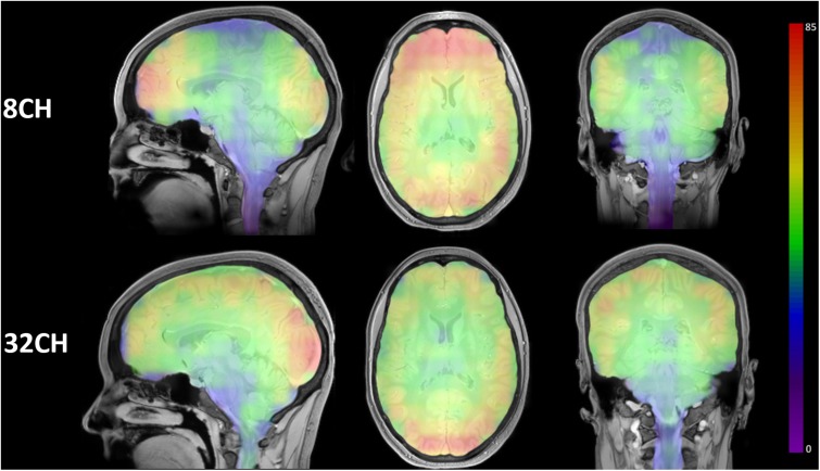
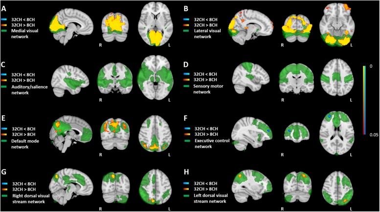
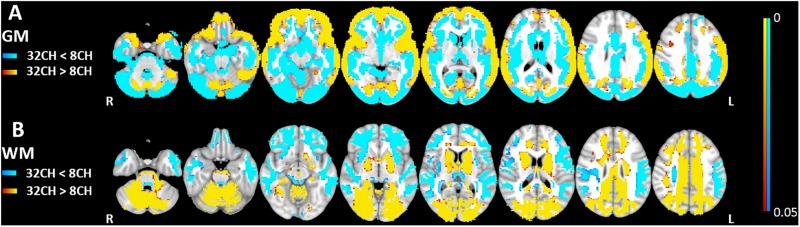
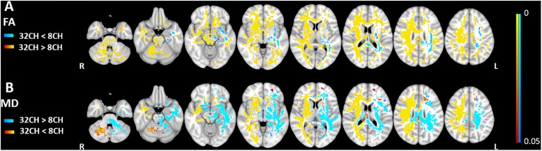
Neuroimaging MRI data in scientific research is increasingly pooled, but the reliability of such studies may be hampered by the use of different hardware elements. This might introduce bias, for example when cross-sectional studies pool data acquired with different head coils, or when longitudinal clinical studies change head coils halfway. In the present study, we aimed to estimate this possible bias introduced by using different head coils to create awareness and to avoid misinterpretation of results. We acquired, with both an 8 channel and 32 channel head coil, T1-weighted, diffusion tensor imaging and resting state fMRI images at 3T MRI (Philips Achieva) with stable acquisition parameters in a large group of cognitively healthy participants ( = 77). Standard analysis methods, i.e., voxel-based morphometry, tract-based spatial statistics and resting state functional network analyses, were used in a within-subject design to compare 8 and 32 channel head coil data. Signal-to-noise ratios (SNR) for both head coils showed similar ranges, although the 32 channel SNR profile was more homogeneous. Our data demonstrates specific patterns of gray and white matter volume differences between head coils (relative volume change of 6 to 9%), related to altered image contrast and therefore, altered tissue segmentation. White matter connectivity (fractional anisotropy and diffusivity measures) showed hemispherical dependent differences between head coils (relative connectivity change of 4 to 6%), and functional connectivity in resting state networks was higher using the 32 channel head coil in posterior cortical areas (relative change up to 27.5%). This study shows that, even when acquisition protocols are harmonized, the results of standardized analysis models can be severely affected by the use of different head coils. Researchers should be aware of this when combining multiple neuroimaging MRI datasets, to prevent coil-related bias and avoid misinterpretation of their findings.
在科学研究中,神经影像学的磁共振成像(MRI)数据越来越多地被汇总,但此类研究的可靠性可能会因使用不同的硬件元件而受到影响。这可能会引入偏差,例如在横断面研究汇总用不同头部线圈采集的数据时,或者纵向临床研究中途更换头部线圈时。在本研究中,我们旨在评估使用不同头部线圈可能引入的这种偏差,以提高认识并避免对结果的错误解读。我们使用8通道和32通道头部线圈,在3T MRI(飞利浦Achieva)上,对一大组认知健康的参与者(n = 77)以稳定的采集参数采集了T1加权、扩散张量成像和静息态功能磁共振成像图像。在受试者内设计中,使用标准分析方法,即基于体素的形态计量学、基于纤维束的空间统计学和静息态功能网络分析,来比较8通道和32通道头部线圈数据。两个头部线圈的信噪比(SNR)显示出相似的范围,尽管32通道的SNR分布更均匀。我们的数据显示了头部线圈之间灰质和白质体积差异的特定模式(相对体积变化为6%至9%),这与图像对比度的改变有关,因此也与组织分割的改变有关。白质连接性(分数各向异性和扩散率测量)显示头部线圈之间存在半球依赖性差异(相对连接性变化为4%至6%),并且在静息态网络中,使用32通道头部线圈时后皮质区域的功能连接性更高(相对变化高达27.5%)。这项研究表明,即使采集方案统一,标准化分析模型的结果也可能会受到使用不同头部线圈的严重影响。研究人员在合并多个神经影像学MRI数据集时应意识到这一点,以防止与线圈相关的偏差并避免对其研究结果的错误解读。