Wang Jing, Yi Xiaoping, Fu Yan, Pang Peipei, Deng Huihuang, Tang Haiyun, Han Zaide, Li Haiping, Nie Jilin, Gong Guanghui, Hu Zhongliang, Tan Zeming, Chen Bihong T
Department of Radiology, Xiangya Hospital, Central South University, Changsha, China.
National Clinical Research Center for Geriatric Disorders, Changsha, China.
Front Oncol. 2021 Oct 27;11:769188. doi: 10.3389/fonc.2021.769188. eCollection 2021.
Early recurrence of glioblastoma after standard treatment makes patient care challenging. This study aimed to assess preoperative magnetic resonance imaging (MRI) radiomics for predicting early recurrence of glioblastoma.
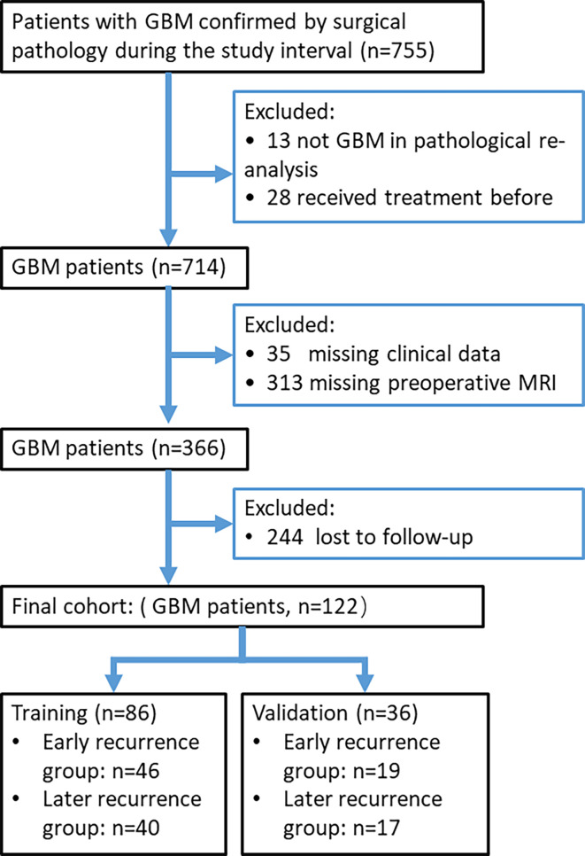
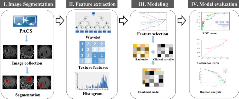
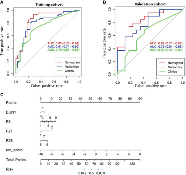
A total of 122 patients (training cohort: n = 86; validation cohort: n = 36) with pathologically confirmed glioblastoma were included in this retrospective study. Preoperative brain MRI images were analyzed for both radiomics and the Visually Accessible Rembrandt Image (VASARI) features of glioblastoma. Models incorporating MRI radiomics, the VASARI parameters, and clinical variables were developed and presented in a nomogram. Performance was assessed based on calibration, discrimination, and clinical usefulness.
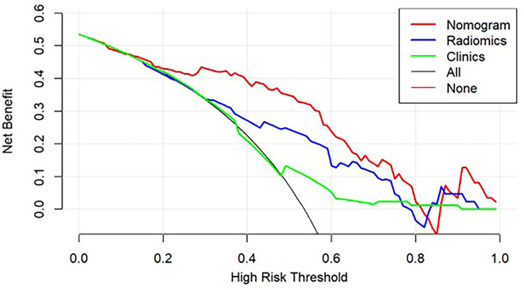
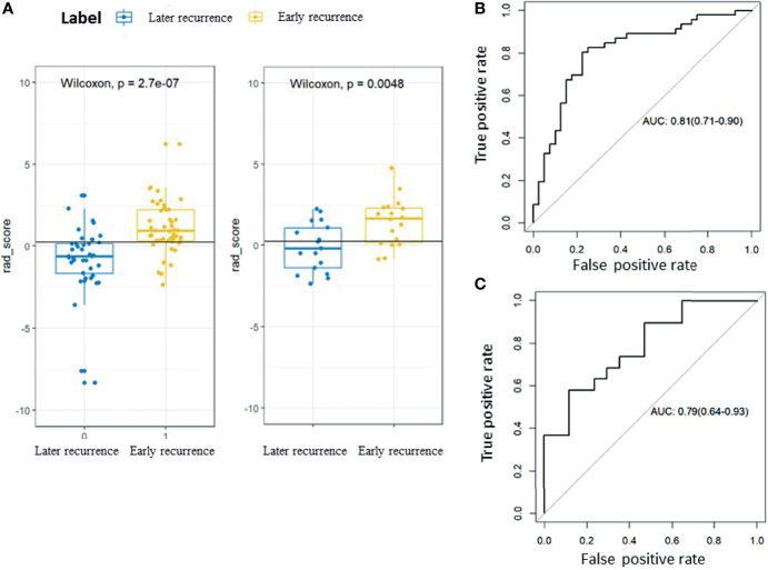
The nomogram consisting of the radiomic signatures, the VASARI parameters, and blood urea nitrogen (BUN) values showed good discrimination between the patients with early recurrence and those with later recurrence, with an area under the curve of 0.85 (95% CI, 0.77-0.94) in the training cohort and 0.84 [95% CI, 0.71-0.97] in the validation cohort. Decision curve analysis demonstrated favorable clinical application of the nomogram.
This study showed the potential usefulness of preoperative brain MRI radiomics in predicting the early recurrence of glioblastoma, which should be helpful in personalized management of glioblastoma.
胶质母细胞瘤在标准治疗后早期复发给患者护理带来挑战。本研究旨在评估术前磁共振成像(MRI)的放射组学特征以预测胶质母细胞瘤的早期复发。
本回顾性研究纳入了122例经病理证实的胶质母细胞瘤患者(训练队列:n = 86;验证队列:n = 36)。对术前脑MRI图像进行放射组学分析以及胶质母细胞瘤的视觉可及性Rembrandt图像(VASARI)特征分析。构建了包含MRI放射组学特征、VASARI参数和临床变量的模型,并以列线图形式呈现。基于校准、鉴别能力和临床实用性对模型性能进行评估。
由放射组学特征、VASARI参数和血尿素氮(BUN)值组成的列线图在早期复发患者和晚期复发患者之间显示出良好的鉴别能力,训练队列中的曲线下面积为0.85(95%CI,0.77 - 0.94),验证队列中的曲线下面积为0.84[95%CI,0.71 - 0.97]。决策曲线分析表明该列线图具有良好的临床应用价值。
本研究表明术前脑MRI放射组学在预测胶质母细胞瘤早期复发方面具有潜在的应用价值,这有助于胶质母细胞瘤的个体化管理。