You Wei, Mao Yitao, Jiao Xiao, Wang Dongcui, Liu Jianling, Lei Peng, Liao Weihua
Department of Radiology, Xiangya Hospital, Central South University, Changsha, China.
National Engineering Research Center of Personalized Diagnostic and Therapeutic Technology, Xiangya Hospital, Central South University, Changsha, China.
Front Oncol. 2023 Mar 22;13:1083216. doi: 10.3389/fonc.2023.1083216. eCollection 2023.
Radiomics features and The Visually AcceSAble Rembrandt Images (VASARI) standard appear to be quantitative and qualitative evaluations utilized to determine glioma grade. This study developed a preoperative model to predict glioma grade and improve the efficacy of clinical strategies by combining these two assessment methods.
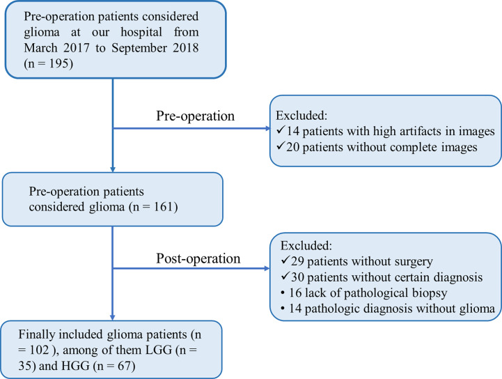
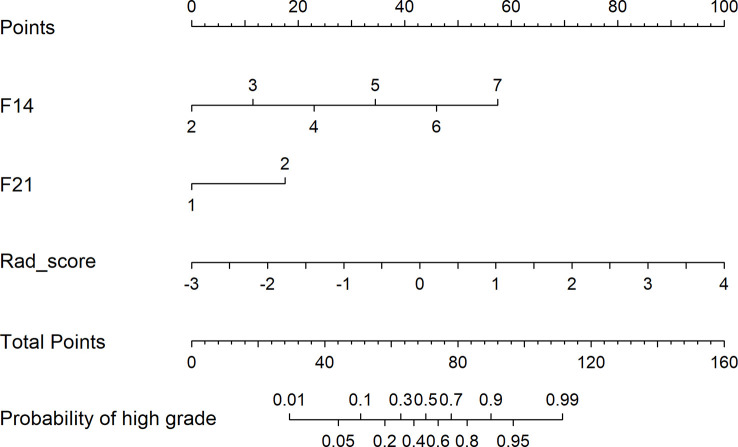
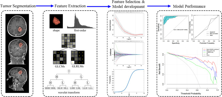
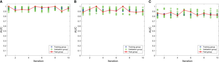
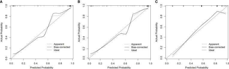
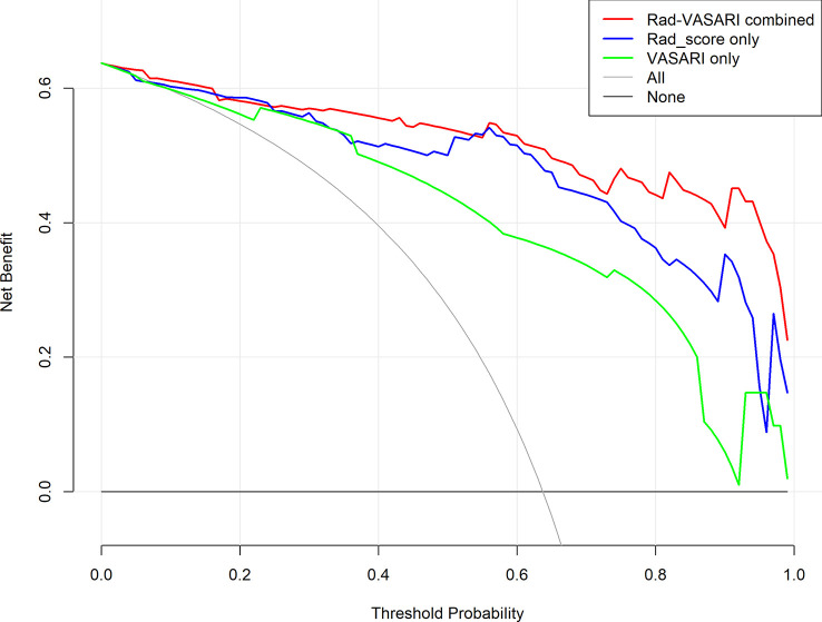
Patients diagnosed with glioma between March 2017 and September 2018 who underwent surgery and histopathology were enrolled in this study. A total of 3840 radiomic features were calculated; however, using the least absolute shrinkage and selection operator (LASSO) method, only 16 features were chosen to generate a radiomic signature. Three predictive models were developed using radiomic features and VASARI standard. The performance and validity of models were evaluated using decision curve analysis and 10-fold nested cross-validation.
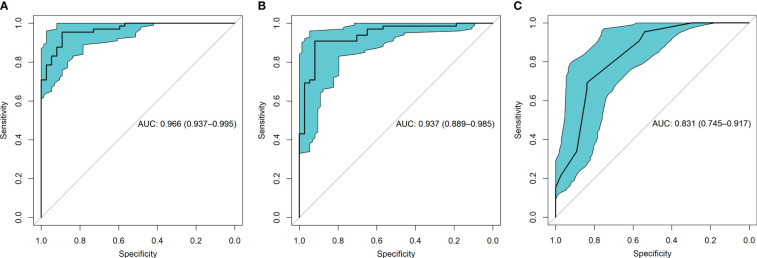
Our study included 102 patients: 35 with low-grade glioma (LGG) and 67 with high-grade glioma (HGG). Model 1 utilized both radiomics and the VASARI standard, which included radiomic signatures, proportion of edema, and deep white matter invasion. Models 2 and 3 were constructed with radiomics or VASARI, respectively, with an area under the receiver operating characteristic curve (AUC) of 0.937 and 0.831, respectively, which was less than that of Model 1, with an AUC of 0.966.
The combination of radiomics features and the VASARI standard is a robust model for predicting glioma grades.
放射组学特征和可视可及的伦勃朗图像(VASARI)标准似乎是用于确定胶质瘤分级的定量和定性评估方法。本研究通过结合这两种评估方法,开发了一种术前模型来预测胶质瘤分级并提高临床策略的疗效。
纳入2017年3月至2018年9月期间接受手术及组织病理学检查且被诊断为胶质瘤的患者。共计算了3840个放射组学特征;然而,使用最小绝对收缩和选择算子(LASSO)方法,仅选择了16个特征来生成放射组学特征标签。使用放射组学特征和VASARI标准开发了三种预测模型。使用决策曲线分析和10倍嵌套交叉验证对模型的性能和有效性进行评估。
我们的研究纳入了102例患者:35例低级别胶质瘤(LGG)患者和67例高级别胶质瘤(HGG)患者。模型1同时使用了放射组学和VASARI标准,包括放射组学特征标签、水肿比例和深部白质侵犯。模型2和模型3分别由放射组学或VASARI构建,其受试者操作特征曲线(AUC)下面积分别为0.937和0.831,均低于模型1的0.966。
放射组学特征与VASARI标准的结合是预测胶质瘤分级的一种可靠模型。