Department of Hand Surgery, China-Japan Union Hospital of Jilin University, 126 Xiantai Avenue, Changchun, 130033, Jilin, China.
Department of Orthopedics, China-Japan Union Hospital of Jilin University, 126 Xiantai Avenue, Changchun, 130033, Jilin, China.
Sci Rep. 2021 Nov 25;11(1):22955. doi: 10.1038/s41598-021-02448-z.
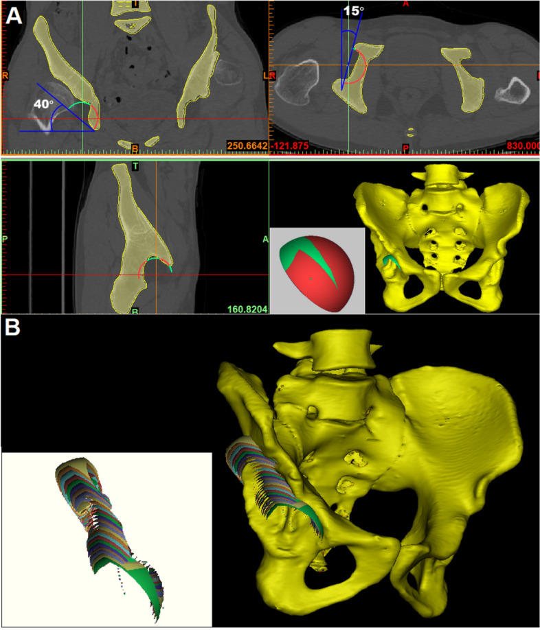
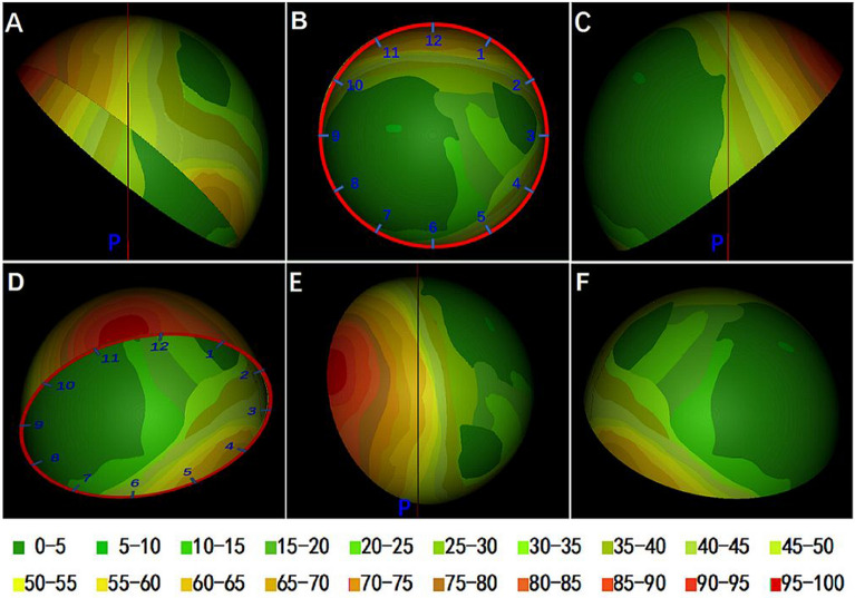
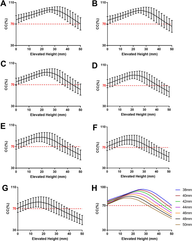
The high hip center technique (HHC) is considered to be feasible for acetabular reconstruction in patients with DDH, but there is little in-depth study of its specific impact on Crowe type II and III DDH. The purpose of this study was to simultaneously analyze the effect of HHC on bone coverage of the cup (CC) in the acetabular reconstruction of type II and III DDH patients and to propose a map of acetabular bone defects from the perspective of the cup. Forty-nine hip CT data of 39 patients with DDH (Crowe type II and III) were collected to simulate acetabular reconstruction by cup models of different sizes (diameter 38mm-50 mm, 2 mm increment) with the HHC technique. The frequency distribution was plotted by overlapping the portions of the 44 mm cups that were not covered by the host bone. The mean CC of cups with sizes of 38 mm, 40 mm, 42 mm, 44 mm, 46 mm, 48 mm, and 50 mm at the true acetabula were 77.85%, 76.71%, 75.73%, 74.56%, 73.68%, 72.51%, and 71.75%, respectively, and the maximum CC increments were 21.24%, 21.58%, 20.86%, 20.04%, 18.62%, 17.18%, and 15.42% (P < 0.001), respectively, after the cups were elevated from the true acetabula. The bone defect map shows that 95% of type II and III DDH acetabula had posterosuperior bone defects, and approximately 60% were located outside the force line of the hip joint. Acetabular cups can meet a CC of more than 70% at the true acetabulum, and approximately 60% of Crowe type II and III DDH patients can obtain satisfactory CC at the true acetabulum by using a 44-mm cup without additional operations.
高位髋关节中心技术(HHC)被认为在 DDH 患者的髋臼重建中是可行的,但对 Crowe II 型和 III 型 DDH 的具体影响研究甚少。本研究旨在同时分析 HHC 对 Crowe II 型和 III 型 DDH 患者髋臼重建中杯骨覆盖率(CC)的影响,并从杯的角度提出髋臼骨缺损图谱。收集了 39 例 DDH(Crowe II 型和 III 型)患者的 49 个髋关节 CT 数据,通过 HHC 技术模拟使用不同大小(直径 38mm-50mm,每 2mm 递增)的杯模型进行髋臼重建。通过重叠宿主骨未覆盖的 44mm 杯的部分,绘制频率分布图。在真髋臼中,38mm、40mm、42mm、44mm、46mm、48mm 和 50mm 杯的真实 CC 分别为 77.85%、76.71%、75.73%、74.56%、73.68%、72.51%和 71.75%,杯从真髋臼抬起后,CC 的最大增量分别为 21.24%、21.58%、20.86%、20.04%、18.62%、17.18%和 15.42%(P<0.001)。骨缺损图谱显示,95%的 Crowe II 型和 III 型 DDH 髋臼存在后上骨缺损,约 60%位于髋关节力线以外。髋臼杯在真髋臼处可获得超过 70%的 CC,约 60%的 Crowe II 型和 III 型 DDH 患者无需额外手术即可通过使用 44mm 杯在真髋臼处获得满意的 CC。