• 文献检索
  • 文档翻译
  • 深度研究
  • 学术资讯
  • Suppr Zotero 插件Zotero 插件
  • 邀请有礼
  • 套餐&价格
  • 历史记录
应用&插件
Suppr Zotero 插件Zotero 插件浏览器插件Mac 客户端Windows 客户端微信小程序
定价
高级版会员购买积分包购买API积分包
服务
文献检索文档翻译深度研究API 文档MCP 服务
关于我们
关于 Suppr公司介绍联系我们用户协议隐私条款
关注我们

Suppr 超能文献

核心技术专利:CN118964589B侵权必究
粤ICP备2023148730 号-1Suppr @ 2026

文献检索

告别复杂PubMed语法,用中文像聊天一样搜索,搜遍4000万医学文献。AI智能推荐,让科研检索更轻松。

立即免费搜索

文件翻译

保留排版,准确专业,支持PDF/Word/PPT等文件格式,支持 12+语言互译。

免费翻译文档

深度研究

AI帮你快速写综述,25分钟生成高质量综述,智能提取关键信息,辅助科研写作。

立即免费体验

并非所有体素都是平等的:使用组织加权均值减少区域 NODDI 指标的估计偏差。

Not all voxels are created equal: Reducing estimation bias in regional NODDI metrics using tissue-weighted means.

机构信息

Centre for Medical Image Computing, Department of Computer Science, University College London, 90 High Holborn, Floor 1, London WC1V6LJ, United Kingdom.

The Dementia Research Centre, Department of Neurodegenerative Disease, UCL Queen Square Institute of Neurology, UCL, London, United Kingdom; UK Dementia Research Institute at UCL, UCL, London, United Kingdom.

出版信息

Neuroimage. 2021 Dec 15;245:118749. doi: 10.1016/j.neuroimage.2021.118749. Epub 2021 Nov 28.

DOI:10.1016/j.neuroimage.2021.118749
PMID:34852276
原文链接:https://pmc.ncbi.nlm.nih.gov/articles/PMC8752961/
Abstract

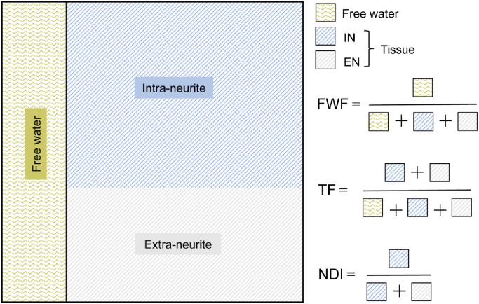
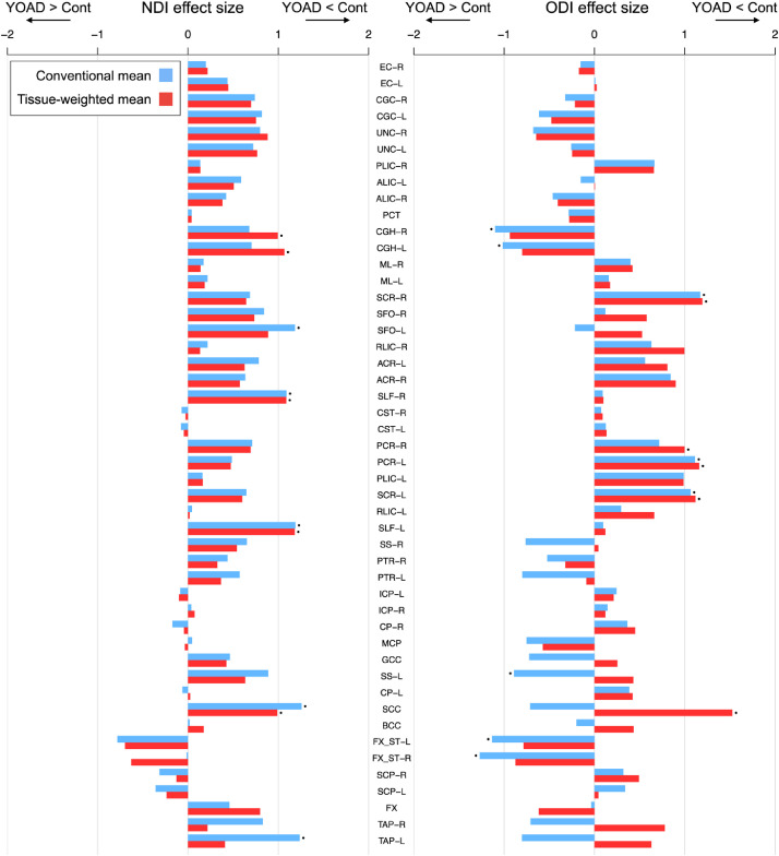
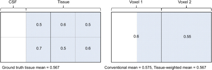
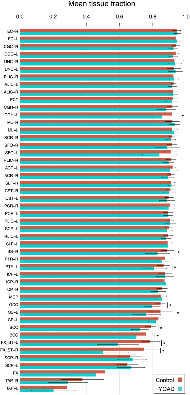
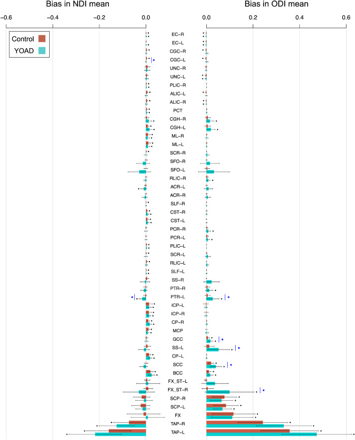
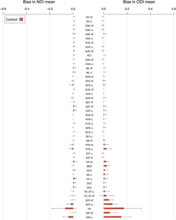
Neurite orientation dispersion and density imaging (NODDI) estimates microstructural properties of brain tissue relating to the organisation and processing capacity of neurites, which are essential elements for neuronal communication. Descriptive statistics of NODDI tissue metrics are commonly analyzed in regions-of-interest (ROI) to identify brain-phenotype associations. Here, the conventional method to calculate the ROI mean weights all voxels equally. However, this produces biased estimates in the presence of CSF partial volume. This study introduces the tissue-weighted mean, which calculates the mean NODDI metric across the tissue within an ROI, utilising the tissue fraction estimate from NODDI to reduce estimation bias. We demonstrate the proposed mean in a study of white matter abnormalities in young onset Alzheimer's disease (YOAD). Results show the conventional mean induces significant bias that correlates with CSF partial volume, primarily affecting periventricular regions and more so in YOAD subjects than in healthy controls. Due to the differential extent of bias between healthy controls and YOAD subjects, the conventional mean under- or over-estimated the effect size for group differences in many ROIs. This demonstrates the importance of using the correct estimation procedure when inferring group differences in studies where the extent of CSF partial volume differs between groups. These findings are robust across different acquisition and processing conditions. Bias persists in ROIs at higher image resolution, as demonstrated using data obtained from the third phase of the Alzheimer's disease neuroimaging initiative (ADNI); and when performing ROI analysis in template space. This suggests that conventional ROI means of NODDI metrics are biased estimates under most contemporary experimental conditions, the correction of which requires the proposed tissue-weighted mean. The tissue-weighted mean produces accurate estimates of ROI means and group differences when ROIs contain voxels with CSF partial volume. In addition to NODDI, the technique can be applied to other multi-compartment models that account for CSF partial volume, such as the free water elimination method. We expect the technique to help generate new insights into normal and abnormal variation in tissue microstructure of regions typically confounded by CSF partial volume, such as those in individuals with larger ventricles due to atrophy associated with neurodegenerative disease.

摘要

神经突方向分散和密度成像(NODDI)估计与神经突的组织和处理能力有关的脑组织的微观结构特性,而神经突是神经元通讯的基本要素。通常在感兴趣区域(ROI)中分析 NODDI 组织指标的描述性统计信息,以识别脑表型关联。在这里,计算 ROI 均值的常规方法是平等地对所有体素进行加权。然而,在存在 CSF 部分体积的情况下,这会产生有偏差的估计。本研究引入了组织加权均值,它利用 NODDI 中的组织分数估计值,在 ROI 内计算组织内的平均 NODDI 指标,以减少估计偏差。我们在青年发病阿尔茨海默病(YOAD)的白质异常研究中展示了所提出的均值。结果表明,常规均值会产生与 CSF 部分体积相关的显著偏差,主要影响脑室周围区域,在 YOAD 患者中比在健康对照组中更为明显。由于健康对照组和 YOAD 患者之间的偏差程度不同,因此在许多 ROI 中,常规均值低估或高估了组间差异的效应大小。这表明,在 CSF 部分体积在组间存在差异的研究中,当推断组间差异时,使用正确的估计程序非常重要。这些发现是稳健的,适用于不同的采集和处理条件。在更高的图像分辨率的 ROI 中仍然存在偏差,正如使用阿尔茨海默病神经影像学倡议(ADNI)第三阶段获得的数据所证明的那样;并且当在模板空间中进行 ROI 分析时也是如此。这表明,在大多数当代实验条件下,NODDI 指标的常规 ROI 均值是有偏差的估计,需要使用所提出的组织加权均值进行校正。当 ROI 包含 CSF 部分体积的体素时,组织加权均值可以产生准确的 ROI 均值和组间差异的估计。除了 NODDI 之外,该技术还可以应用于其他考虑 CSF 部分体积的多腔室模型,例如自由水消除法。我们希望该技术有助于深入了解通常受 CSF 部分体积混淆的区域的组织微观结构的正常和异常变化,例如由于与神经退行性疾病相关的萎缩导致脑室较大的个体的区域。

https://cdn.ncbi.nlm.nih.gov/pmc/blobs/ffd2/8752961/ea30a05e920b/gr6.jpg
https://cdn.ncbi.nlm.nih.gov/pmc/blobs/ffd2/8752961/f4735c4d667a/gr1.jpg
https://cdn.ncbi.nlm.nih.gov/pmc/blobs/ffd2/8752961/d9311db0a2dc/gr2.jpg
https://cdn.ncbi.nlm.nih.gov/pmc/blobs/ffd2/8752961/dabc21d95123/gr3.jpg
https://cdn.ncbi.nlm.nih.gov/pmc/blobs/ffd2/8752961/c52d37265516/gr4.jpg
https://cdn.ncbi.nlm.nih.gov/pmc/blobs/ffd2/8752961/ba96421e26d0/gr5.jpg
https://cdn.ncbi.nlm.nih.gov/pmc/blobs/ffd2/8752961/ea30a05e920b/gr6.jpg

相似文献

1
Not all voxels are created equal: Reducing estimation bias in regional NODDI metrics using tissue-weighted means.并非所有体素都是平等的:使用组织加权均值减少区域 NODDI 指标的估计偏差。
Neuroimage. 2021 Dec 15;245:118749. doi: 10.1016/j.neuroimage.2021.118749. Epub 2021 Nov 28.
2
MTE-NODDI: Multi-TE NODDI for disentangling non-T2-weighted signal fractions from compartment-specific T2 relaxation times.MTE-NODDI:用于从具有特定隔室的 T2 弛豫时间中分离非 T2 加权信号分数的多 TE NODDI。
Neuroimage. 2020 Aug 15;217:116906. doi: 10.1016/j.neuroimage.2020.116906. Epub 2020 May 7.
3
Age effects and sex differences in human brain white matter of young to middle-aged adults: A DTI, NODDI, and q-space study.青年至中年成年人脑白质的年龄效应和性别差异:一项扩散张量成像(DTI)、神经突方向离散与密度成像(NODDI)和q空间研究
Neuroimage. 2016 Mar;128:180-192. doi: 10.1016/j.neuroimage.2015.12.033. Epub 2015 Dec 24.
4
Microstructural White Matter Alterations in Mild Cognitive Impairment and Alzheimer's Disease : Study Based on Neurite Orientation Dispersion and Density Imaging (NODDI).轻度认知障碍和阿尔茨海默病的微观结构白质改变:基于神经丝取向分散和密度成像(NODDI)的研究。
Clin Neuroradiol. 2020 Sep;30(3):569-579. doi: 10.1007/s00062-019-00805-0. Epub 2019 Jun 7.
5
Longitudinal Reproducibility of Neurite Orientation Dispersion and Density Imaging (NODDI) Derived Metrics in the White Matter.神经丝取向分散和密度成像(NODDI)衍生指标在白质中的纵向可重复性。
Neuroscience. 2021 Mar 1;457:165-185. doi: 10.1016/j.neuroscience.2021.01.005. Epub 2021 Jan 17.
6
ApoE influences regional white-matter axonal density loss in Alzheimer's disease.载脂蛋白E影响阿尔茨海默病中区域白质轴突密度的丧失。
Neurobiol Aging. 2017 Sep;57:8-17. doi: 10.1016/j.neurobiolaging.2017.04.021. Epub 2017 May 3.
7
Neurite density imaging versus imaging of microscopic anisotropy in diffusion MRI: A model comparison using spherical tensor encoding.扩散磁共振成像中神经突密度成像与微观各向异性成像的比较:使用球张量编码的模型对比
Neuroimage. 2017 Feb 15;147:517-531. doi: 10.1016/j.neuroimage.2016.11.053. Epub 2016 Nov 27.
8
Application of neurite orientation dispersion and density imaging to characterize brain microstructural abnormalities in type-2 diabetics with mild cognitive impairment.神经突方向分散和密度成像在 2 型糖尿病伴轻度认知障碍患者脑微结构异常中的应用。
J Magn Reson Imaging. 2019 Sep;50(3):889-898. doi: 10.1002/jmri.26687. Epub 2019 Feb 19.
9
Applicability of multiple quantitative magnetic resonance methods in genetic brain white matter disorders.多种定量磁共振方法在遗传性脑白质疾病中的适用性
J Neuroimaging. 2024 Jan-Feb;34(1):61-77. doi: 10.1111/jon.13167. Epub 2023 Nov 4.
10
Bingham-NODDI: Mapping anisotropic orientation dispersion of neurites using diffusion MRI.宾厄姆-神经突方向离散度成像(Bingham-NODDI):利用扩散磁共振成像绘制神经突的各向异性方向离散度图。
Neuroimage. 2016 Jun;133:207-223. doi: 10.1016/j.neuroimage.2016.01.046. Epub 2016 Jan 28.

引用本文的文献

1
Evaluation of limbic microstructural abnormalities in temporal lobe epilepsy: A neurite orientation distribution and density imaging study.颞叶癫痫边缘系统微观结构异常的评估:一项神经突方向分布与密度成像研究。
Epilepsia. 2025 Jun 4. doi: 10.1111/epi.18488.
2
Cortical microstructural abnormalities in dementia with Lewy bodies and their associations with Alzheimer's disease copathologies.路易体痴呆中的皮质微结构异常及其与阿尔茨海默病共病的关联。
NPJ Parkinsons Dis. 2025 May 12;11(1):124. doi: 10.1038/s41531-025-00944-x.
3
White matter characterization in regions of edema surrounding meningioma brain tumor using diffusion MRI: A comparative study of DTI and NODDI.

本文引用的文献

1
The evolution of white matter microstructural changes after mild traumatic brain injury: A longitudinal DTI and NODDI study.轻度创伤性脑损伤后白质微观结构变化的演变:一项纵向扩散张量成像(DTI)和神经突方向离散与密度成像(NODDI)研究
Sci Adv. 2020 Aug 7;6(32):eaaz6892. doi: 10.1126/sciadv.aaz6892. eCollection 2020 Aug.
2
Biological and clinical characteristics of gene carriers far from predicted onset in the Huntington's disease Young Adult Study (HD-YAS): a cross-sectional analysis.亨廷顿病青年研究(HD-YAS)中基因携带者的生物学和临床特征:一项横断面分析。
Lancet Neurol. 2020 Jun;19(6):502-512. doi: 10.1016/S1474-4422(20)30143-5. Epub 2020 May 26.
3
使用扩散磁共振成像对脑膜瘤脑肿瘤周围水肿区域的白质特征进行研究:扩散张量成像(DTI)与神经突方向离散与密度成像(NODDI)的对比研究
medRxiv. 2025 Apr 8:2025.04.07.25325393. doi: 10.1101/2025.04.07.25325393.
4
Associations among white matter microstructural changes and the development of emotional reactivity and regulation in infancy.白质微观结构变化与婴儿期情绪反应性和调节能力发展之间的关联。
Mol Psychiatry. 2025 Apr 23. doi: 10.1038/s41380-025-03025-w.
5
An Investigation of Corticospinal Tract Microstructural Integrity in ARSACS Using a Profilometry MRI Analysis: Results From the PROSPAX Study.使用轮廓测量MRI分析对ARSACS患者皮质脊髓束微结构完整性的研究:PROSPAX研究结果
Eur J Neurol. 2025 Apr;32(4):e70128. doi: 10.1111/ene.70128.
6
Lifespan Normative Models of White Matter Fractional Anisotropy: Applications to Early Psychosis.白质分数各向异性的寿命规范性模型:在早期精神病中的应用。
bioRxiv. 2024 Dec 11:2024.12.11.627897. doi: 10.1101/2024.12.11.627897.
7
Quantitative MRI distinguishes different leukodystrophies and correlates with clinical measures.定量磁共振成像可区分不同的脑白质营养不良,并与临床指标相关。
Eur Radiol. 2025 Apr;35(4):1845-1857. doi: 10.1007/s00330-024-11089-5. Epub 2024 Sep 25.
8
An MRI evaluation of white matter involvement in paradigmatic forms of spastic ataxia: results from the multi-center PROSPAX study.MRI 评估痉挛性共济失调典型形式的脑白质受累:多中心 PROSPAX 研究结果。
J Neurol. 2024 Aug;271(8):5468-5477. doi: 10.1007/s00415-024-12505-y. Epub 2024 Jun 16.
9
Early Infant Prefrontal Cortical Microstructure Predicts Present and Future Emotionality.早期婴儿前额皮质微观结构预测现在和未来的情绪性。
Biol Psychiatry. 2024 Dec 15;96(12):959-970. doi: 10.1016/j.biopsych.2024.04.001. Epub 2024 Apr 9.
10
Feature similarity gradients detect alterations in the neonatal cortex associated with preterm birth.特征相似性梯度可检测与早产相关的新生儿皮层变化。
Hum Brain Mapp. 2024 Mar;45(4):e26660. doi: 10.1002/hbm.26660.
Magnitude and timing of major white matter tract maturation from infancy through adolescence with NODDI.
利用 NODDI 研究从婴儿期到青春期主要白质束的发育程度和时间。
Neuroimage. 2020 May 15;212:116672. doi: 10.1016/j.neuroimage.2020.116672. Epub 2020 Feb 21.
4
MR Biomarkers of Degenerative Brain Disorders Derived From Diffusion Imaging.基于扩散成像的退行性脑疾病的磁共振生物标志物
J Magn Reson Imaging. 2020 Dec;52(6):1620-1636. doi: 10.1002/jmri.27019. Epub 2019 Dec 13.
5
White matter alterations in early-stage Alzheimer's disease: A tract-specific study.早期阿尔茨海默病中的白质改变:一项基于纤维束的研究。
Alzheimers Dement (Amst). 2019 Aug 21;11:576-587. doi: 10.1016/j.dadm.2019.06.003. eCollection 2019 Dec.
6
The spatial correspondence and genetic influence of interhemispheric connectivity with white matter microstructure.大脑两半球间连接的空间对应关系及其与白质微观结构的遗传影响。
Nat Neurosci. 2019 May;22(5):809-819. doi: 10.1038/s41593-019-0379-2. Epub 2019 Apr 15.
7
Neurite orientation and dispersion density imaging (NODDI) detects cortical and corticospinal tract degeneration in ALS.神经突方向分布密度成像(NODDI)可检测 ALS 中的皮质和皮质脊髓束变性。
J Neurol Neurosurg Psychiatry. 2019 Apr;90(4):404-411. doi: 10.1136/jnnp-2018-318830. Epub 2018 Oct 25.
8
Neurite density is reduced in the presymptomatic phase of disease.神经突密度在疾病的无症状前期降低。
J Neurol Neurosurg Psychiatry. 2019 Apr;90(4):387-394. doi: 10.1136/jnnp-2018-318994. Epub 2018 Oct 24.
9
In vivo characterization of white matter pathology in premanifest huntington's disease.在症状前亨廷顿病中白质病理学的体内特征描述。
Ann Neurol. 2018 Oct;84(4):497-504. doi: 10.1002/ana.25309. Epub 2018 Sep 15.
10
Diffusion markers of dendritic density and arborization in gray matter predict differences in intelligence.树突密度和分支的扩散标记物可以预测灰质中的智力差异。
Nat Commun. 2018 May 15;9(1):1905. doi: 10.1038/s41467-018-04268-8.