Laboratorio de Parásitos Anaerobios, Instituto Tecnológico Chascomús (INTECH), Consejo Nacional de Investigaciones Científicas y Técnicas - Universidad Nacional de General San Martín (CONICET-UNSAM), Chascomús, Argentina.
Departamento de Microbiologia, Instituto Aggeu Magalhães, FIOCRUZ, Recife, Brazil.
Front Cell Infect Microbiol. 2021 Nov 9;11:757185. doi: 10.3389/fcimb.2021.757185. eCollection 2021.
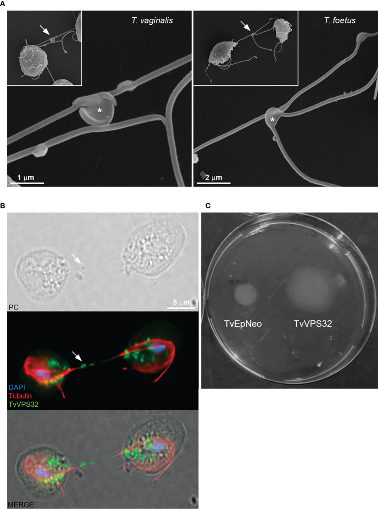
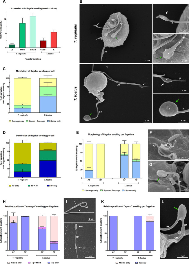
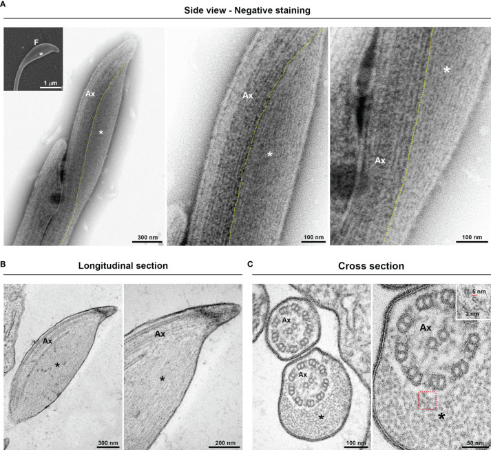
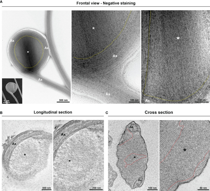
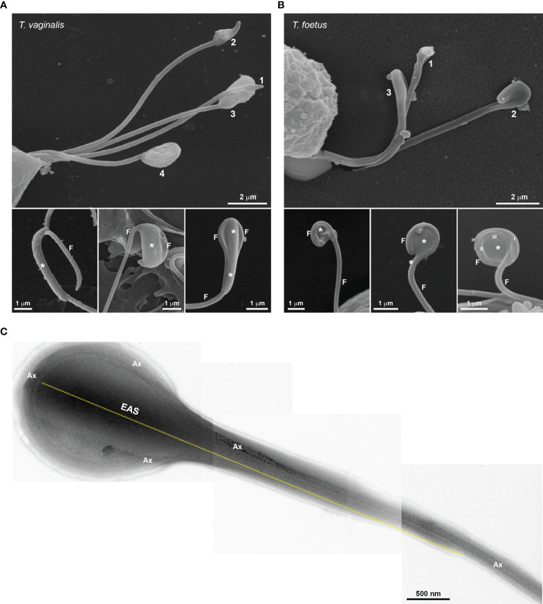
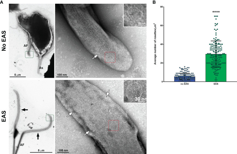
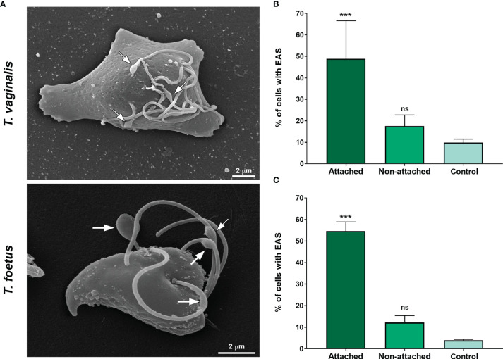
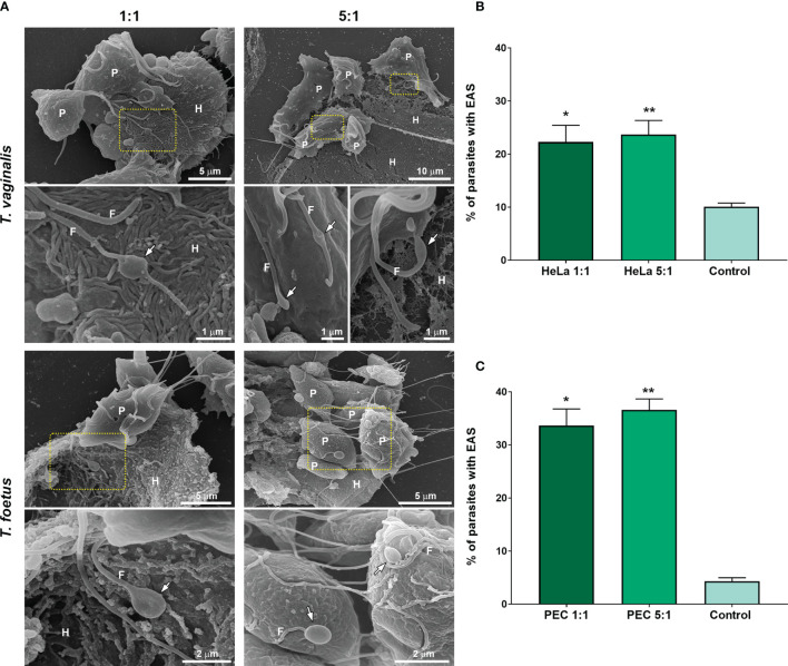
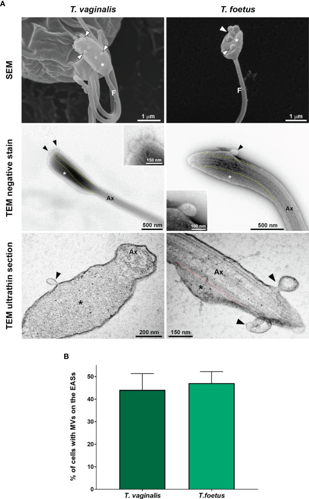
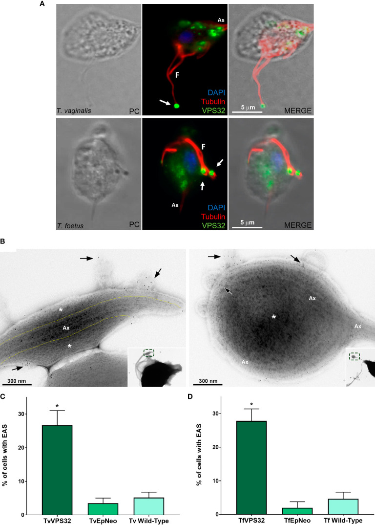
and are extracellular flagellated parasites that inhabit humans and other mammals, respectively. In addition to motility, flagella act in a variety of biological processes in different cell types, and extra-axonemal structures (EASs) have been described as fibrillar structures that provide mechanical support and act as metabolic, homeostatic, and sensory platforms in many organisms. It has been assumed that and do not have EASs. However, here, we used complementary electron microscopy techniques to reveal the ultrastructure of EASs in both parasites. Such EASs are thin filaments (3-5 nm diameter) running longitudinally along the axonemes and surrounded by the flagellar membrane, forming prominent flagellar swellings. We observed that the formation of EAS increases after parasite adhesion on the host cells, fibronectin, and precationized surfaces. A high number of rosettes, clusters of intramembrane particles that have been proposed as sensorial structures, and microvesicles protruding from the membrane were observed in the EASs. Our observations demonstrate that and can connect to themselves by EASs present in flagella. The protein VPS32, a member of the ESCRT-III complex crucial for diverse membrane remodeling events, the pinching off and release of microvesicles, was found in the surface as well as in microvesicles protruding from EASs. Moreover, we demonstrated that the formation of EAS also increases in parasites overexpressing VPS32 and that -VPS32 parasites showed greater motility in semisolid agar. These results provide valuable data about the role of the flagellar EASs in the cell-to-cell communication and pathogenesis of these extracellular parasites.
和 分别是寄生于人类和其他哺乳动物的细胞外鞭毛寄生虫。除了运动性之外,鞭毛在不同细胞类型的多种生物学过程中发挥作用,并且已经描述了额外轴丝结构(EAS)作为提供机械支撑的纤维状结构,并在许多生物体中充当代谢、动态平衡和感觉平台。人们一直认为 和 没有 EAS。然而,在这里,我们使用互补的电子显微镜技术来揭示这两种寄生虫中 EAS 的超微结构。这些 EAS 是沿轴丝纵向运行的细纤维(3-5nm 直径),并被鞭毛膜包围,形成明显的鞭毛肿胀。我们观察到,在寄生虫附着在宿主细胞、纤维连接蛋白和预处理表面后,EAS 的形成增加。在 EAS 中观察到大量的花冠状结构、被提议为感觉结构的膜内颗粒簇以及从膜中突出的微囊泡。我们的观察表明, 和 可以通过鞭毛中的 EAS 相互连接。VPS32 蛋白是 ESCRT-III 复合物的成员,对于多种膜重塑事件、微囊泡的缢缩和释放至关重要,在表面以及从 EAS 中突出的微囊泡中都有发现。此外,我们证明了在过表达 VPS32 的寄生虫中,EAS 的形成也会增加,并且 -VPS32 寄生虫在半固体琼脂中的运动性更强。这些结果提供了有关鞭毛 EAS 在这些细胞外寄生虫的细胞间通讯和发病机制中的作用的有价值的数据。