Xiang Fei, Wei Shumei, Liu Xingyu, Liang Xiaoyuan, Yang Lili, Yan Sheng
Department of Hepatobiliary and Pancreatic Surgery, The Second Affiliated Hospital, Zhejiang University School of Medicine, Hangzhou, China.
Department of Pathology, The Second Affiliated Hospital, Zhejiang University School of Medicine, Hangzhou, China.
Front Oncol. 2021 Nov 19;11:774117. doi: 10.3389/fonc.2021.774117. eCollection 2021.
Microvascular invasion (MVI) has been shown to be closely associated with postoperative recurrence and metastasis in patients with intrahepatic cholangiocarcinoma (ICC). We aimed to develop a radiomics prediction model based on contrast-enhanced CT (CECT) to distinguish MVI in patients with mass-forming ICC.
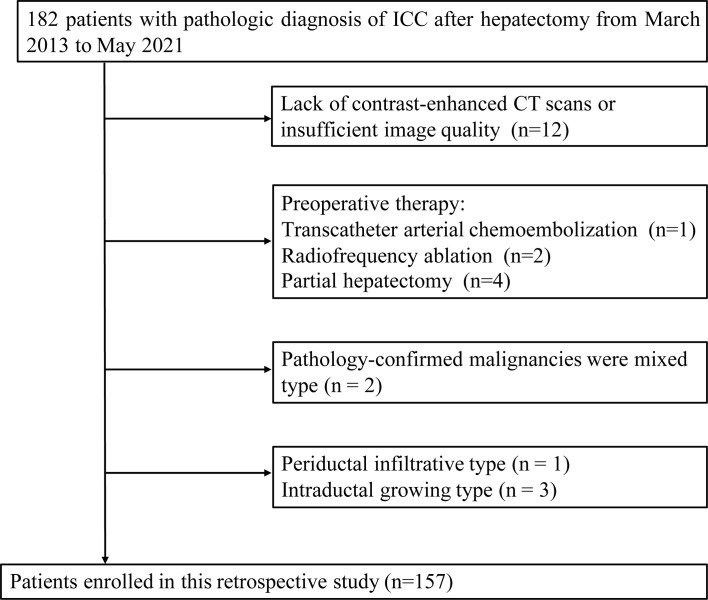
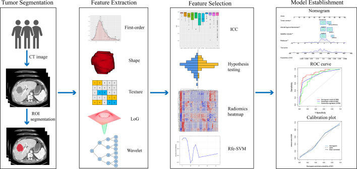
157 patients were included and randomly divided into training (n=110) and test (n=47) datasets. Radiomic signatures were built based on the recursive feature elimination support vector machine (Rfe-SVM) algorithm. Significant clinical-radiologic factors were screened, and a clinical model was built by multivariate logistic regression. A nomogram was developed by integrating radiomics signature and the significant clinical risk factors.
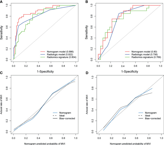
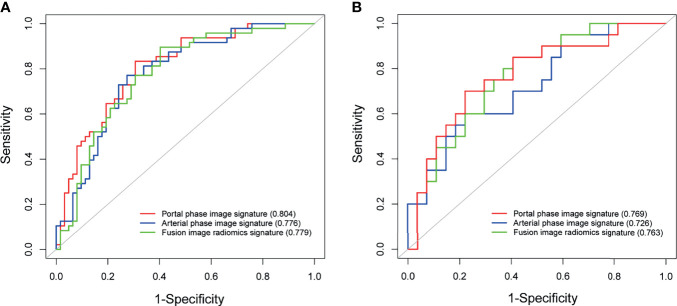
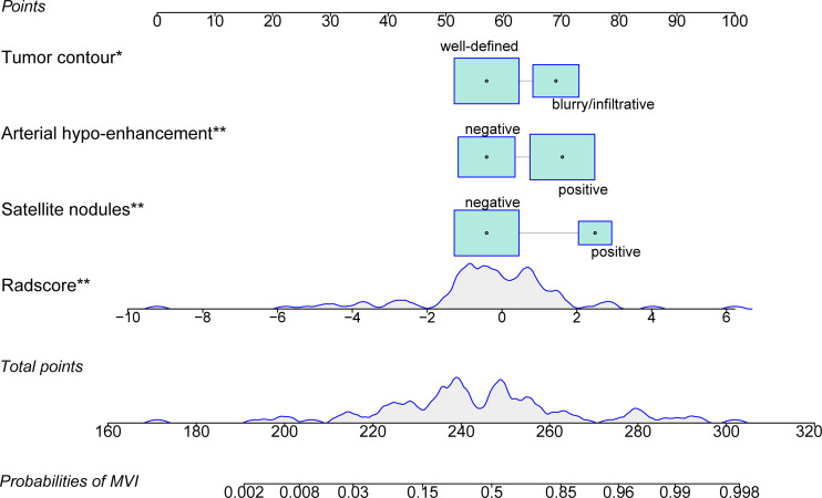
The portal phase image radiomics signature with 6 features was constructed and provided an area under the receiver operating characteristic curve (AUC) of 0.804 in the training and 0.769 in the test datasets. Three significant predictors, including satellite nodules (odds ratio [OR]=13.73), arterial hypo-enhancement (OR=4.31), and tumor contour (OR=4.99), were identified by multivariate analysis. The clinical model using these predictors exhibited an AUC of 0.822 in the training and 0.756 in the test datasets. The nomogram combining significant clinical factors and radiomics signature achieved satisfactory prediction efficacy, showing an AUC of 0.886 in the training and 0.80 in the test datasets.
Both CECT radiomics analysis and radiologic factors have the potential for MVI prediction in mass-forming ICC patients. The nomogram can further improve the prediction efficacy.
微血管侵犯(MVI)已被证明与肝内胆管癌(ICC)患者术后复发和转移密切相关。我们旨在开发一种基于对比增强CT(CECT)的放射组学预测模型,以鉴别肿块型ICC患者中的MVI。
纳入157例患者,随机分为训练集(n = 110)和测试集(n = 47)。基于递归特征消除支持向量机(Rfe-SVM)算法构建放射组学特征。筛选出显著的临床-放射学因素,并通过多因素逻辑回归建立临床模型。通过整合放射组学特征和显著的临床风险因素开发列线图。
构建了具有6个特征的门静脉期图像放射组学特征,在训练集中的受试者工作特征曲线下面积(AUC)为0.804,在测试集中为0.769。多因素分析确定了3个显著预测因素,包括卫星结节(比值比[OR]=13.73)、动脉期低强化(OR = 4.31)和肿瘤轮廓(OR = 4.99)。使用这些预测因素的临床模型在训练集中的AUC为0.822,在测试集中为0.756。结合显著临床因素和放射组学特征的列线图实现了令人满意的预测效能,在训练集中的AUC为0.886,在测试集中为0.80。
CECT放射组学分析和放射学因素在肿块型ICC患者的MVI预测中均具有潜力。列线图可进一步提高预测效能。