Dupin Nicolas, Jary Aude, Boussouar Samia, Syrykh Charlotte, Gandjbakhche Amir, Bergeret Sébastien, Palich Romain
Dermatology Department, Cochin Hospital, AP-HP, Institut Cochin, INSERM 1016, Université de Paris, 75014 Paris, France.
Virology Department, Pitié-Salpêtrière Hospital, AP-HP, Pierre Louis Epidemiology and Public Health Institute (iPLESP), INSERM 1136, Sorbonne University, 75013 Paris, France.
Cancers (Basel). 2021 Nov 25;13(23):5927. doi: 10.3390/cancers13235927.
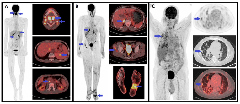
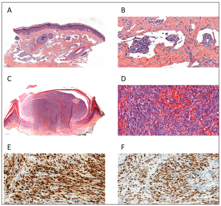
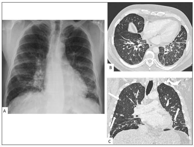
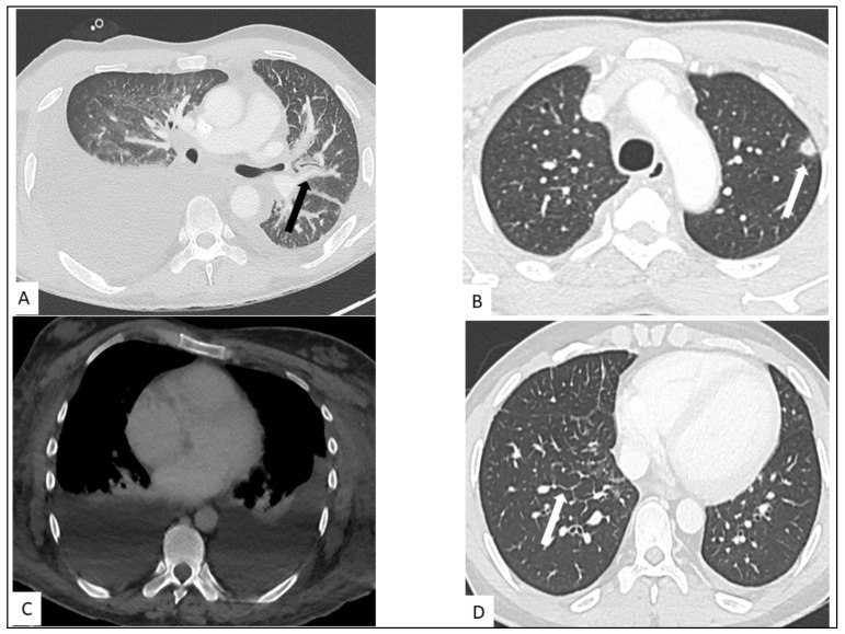
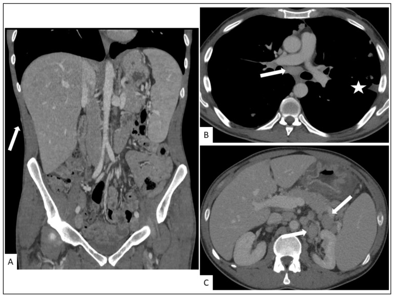
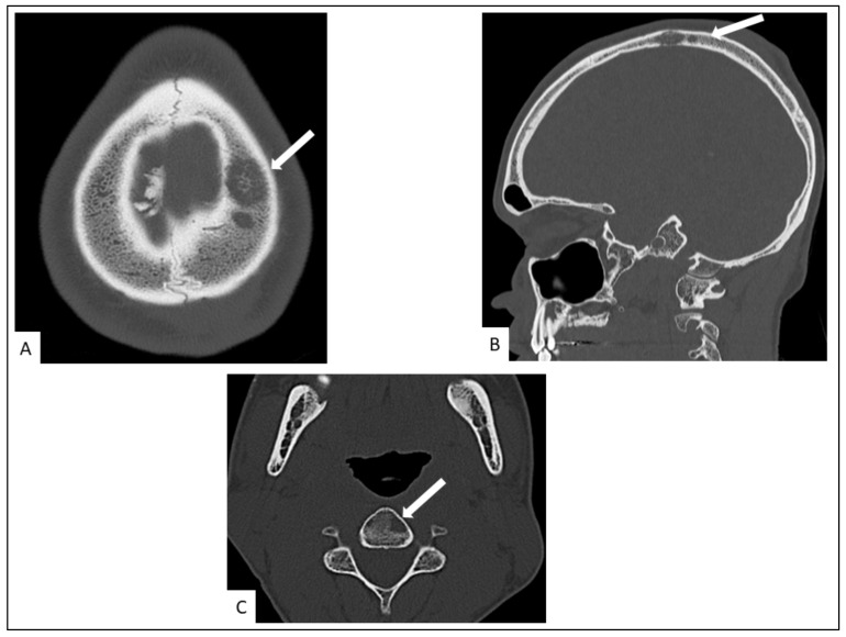
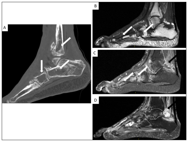
Kaposi's sarcoma (KS) is a rare, atypical malignancy associated with immunosuppression and can be qualified as an opportunistic tumor, which responds to immune modulation or restoration. Four different epidemiological forms have been individualized (AIDS-related, iatrogenic, endemic or classic KS). Although clinical examination is sufficient to diagnose cutaneous lesions of KS, additional explorations are necessary in order to detect lesions involving other organs. New histological markers have been developed in recent years concerning the detection of HHV-8 latent or lytic proteins in the lesions, helping to confirm the diagnosis when it is clinically doubtful. More recently, the evaluation of the local immune response has also been shown to provide some guidance in choosing the appropriate therapeutic option when necessary. We also review the indication and the results of conventional radiological imaging and of non-invasive imaging tools such as F-fluoro-deoxy-glucose positron emission tomography, thermography and laser Doppler imaging for the diagnosis of KS and for the follow-up of therapeutic response in patients requiring systemic treatment.
卡波西肉瘤(KS)是一种与免疫抑制相关的罕见非典型恶性肿瘤,可被视为一种机会性肿瘤,对免疫调节或恢复有反应。已确定有四种不同的流行病学形式(艾滋病相关型、医源性、地方性或经典型KS)。虽然临床检查足以诊断KS的皮肤病变,但为了检测累及其他器官的病变,还需要进行额外的检查。近年来,已开发出新的组织学标志物用于检测病变中HHV-8潜伏或裂解蛋白,在临床诊断存疑时有助于确诊。最近,局部免疫反应的评估也已显示在必要时为选择合适的治疗方案提供一些指导。我们还综述了传统放射影像学以及诸如F-氟脱氧葡萄糖正电子发射断层扫描、热成像和激光多普勒成像等非侵入性成像工具在KS诊断及需要全身治疗患者的治疗反应随访中的应用指征和结果。