PHEniX Laboratory, Department of Pulmonary Medicine, Amsterdam UMC, Vrije Universiteit Amsterdam, Amsterdam Cardiovascular Sciences, 1081 HZ Amsterdam, The Netherlands.
Department of Physiology, Amsterdam UMC, Vrije Universiteit Amsterdam, Amsterdam Cardiovascular Sciences, 1081 HZ Amsterdam, The Netherlands.
Cells. 2021 Dec 20;10(12):3595. doi: 10.3390/cells10123595.
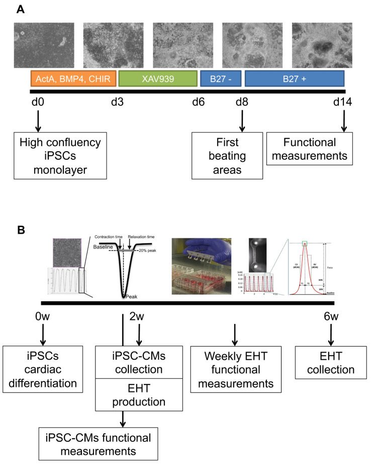
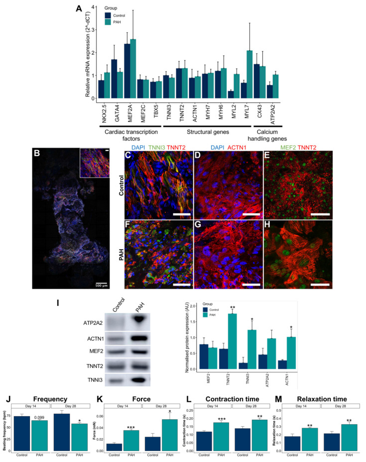
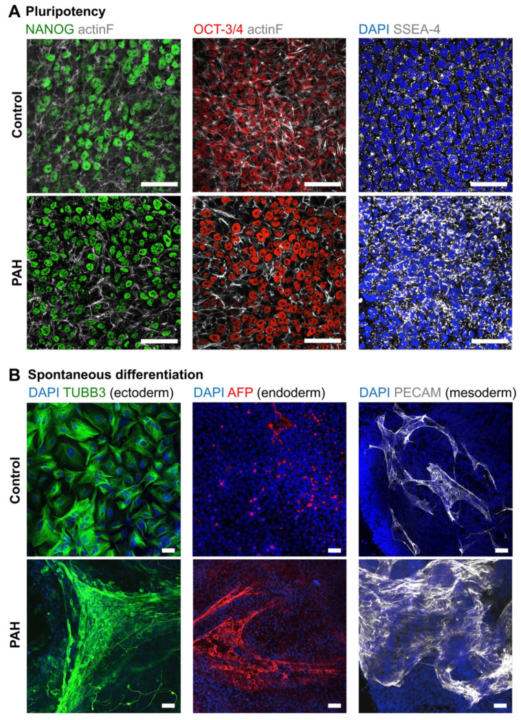
Pulmonary arterial hypertension (PAH) patients eventually die of right heart failure (RHF). Currently, there is no suitable pre-clinical model to study PAH. Therefore, we aim to develop a right heart dysfunction (RHD) model using the 3-dimensional engineered heart tissue (EHT) approach and cardiomyocytes derived from patient-induced pluripotent stem cells (iPSCs) to unravel the mechanisms that determine the fate of a pressure-overloaded right ventricle. iPSCs from PAH and healthy control subjects were differentiated into cardiomyocytes (iPSC-CMs), incorporated into the EHT, and maintained for 28 days. In comparison with control iPSC-CMs, PAH-derived iPSC-CMs exhibited decreased beating frequency and increased contraction and relaxation times. iPSC-CM alignment within the EHT was observed. PAH-derived EHTs exhibited higher force, and contraction and relaxation times compared with control EHTs. Increased afterload was induced using 2× stiffer posts from day 0. Due to high variability, there were no functional differences between normal and stiffer EHTs, and no differences in the hypertrophic gene expression. In conclusion, under baseline spontaneous conditions, PAH-derived iPSC-CMs and EHTs show prolonged contraction compared with controls, as observed clinically in PAH patients. Further optimization of the hypertrophic model and profound characterization may provide a platform for disease modelling and drug screening.
肺动脉高压 (PAH) 患者最终会因右心衰竭 (RHF) 而死亡。目前,尚无合适的临床前模型来研究 PAH。因此,我们旨在使用 3 维工程心脏组织 (EHT) 方法和源自患者诱导多能干细胞 (iPSC) 的心肌细胞来开发右心功能障碍 (RHD) 模型,以揭示决定压力超负荷右心室命运的机制。来自 PAH 和健康对照者的 iPSC 分化为心肌细胞 (iPSC-CMs),并整合到 EHT 中,并维持 28 天。与对照 iPSC-CMs 相比,PAH 来源的 iPSC-CMs 的搏动频率降低,收缩和舒张时间增加。观察到 iPSC-CM 在 EHT 中的排列。与对照 EHT 相比,PAH 来源的 EHT 显示出更高的力、收缩和舒张时间。从第 0 天开始,使用 2 倍更硬的支柱施加更高的后负荷。由于变异性高,正常和更硬的 EHT 之间没有功能差异,也没有肥厚基因表达的差异。总之,在基线自发条件下,与对照组相比,PAH 来源的 iPSC-CMs 和 EHT 显示出延长的收缩,这在 PAH 患者的临床观察中是如此。进一步优化肥大模型和深入表征可能为疾病建模和药物筛选提供一个平台。