• 文献检索
  • 文档翻译
  • 深度研究
  • 学术资讯
  • Suppr Zotero 插件Zotero 插件
  • 邀请有礼
  • 套餐&价格
  • 历史记录
应用&插件
Suppr Zotero 插件Zotero 插件浏览器插件Mac 客户端Windows 客户端微信小程序
定价
高级版会员购买积分包购买API积分包
服务
文献检索文档翻译深度研究API 文档MCP 服务
关于我们
关于 Suppr公司介绍联系我们用户协议隐私条款
关注我们

Suppr 超能文献

核心技术专利:CN118964589B侵权必究
粤ICP备2023148730 号-1Suppr @ 2026

文献检索

告别复杂PubMed语法,用中文像聊天一样搜索,搜遍4000万医学文献。AI智能推荐,让科研检索更轻松。

立即免费搜索

文件翻译

保留排版,准确专业,支持PDF/Word/PPT等文件格式,支持 12+语言互译。

免费翻译文档

深度研究

AI帮你快速写综述,25分钟生成高质量综述,智能提取关键信息,辅助科研写作。

立即免费体验

用于……成像的具有低温聚焦离子束铣削的超分辨率共聚焦冷冻扫描透射电子显微镜联用技术

Super-resolution confocal cryo-CLEM with cryo-FIB milling for imaging of .

作者信息

Sexton Danielle L, Burgold Steffen, Schertel Andreas, Tocheva Elitza I

机构信息

Department of Microbiology and Immunology, University of British Columbia, Vancouver, Canada.

ZEISS Microscopy Customer Center, Oberkochen, Germany.

出版信息

Curr Res Struct Biol. 2021 Dec 13;4:1-9. doi: 10.1016/j.crstbi.2021.12.001. eCollection 2022.

DOI:10.1016/j.crstbi.2021.12.001
PMID:34977598
原文链接:https://pmc.ncbi.nlm.nih.gov/articles/PMC8688812/
Abstract

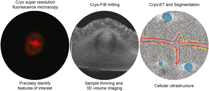
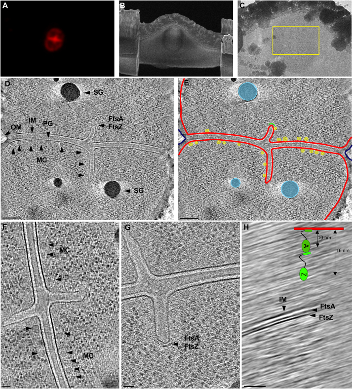
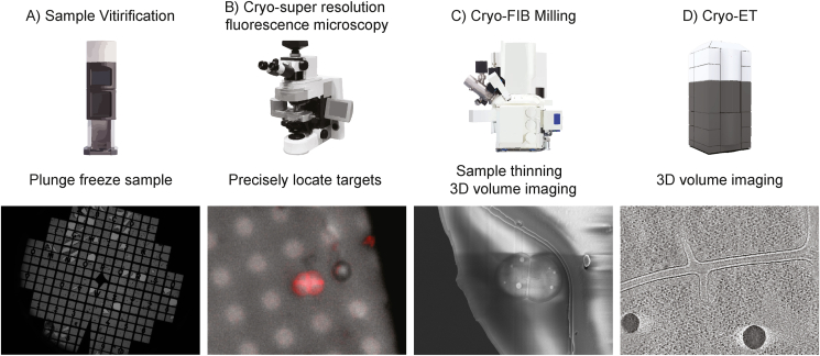
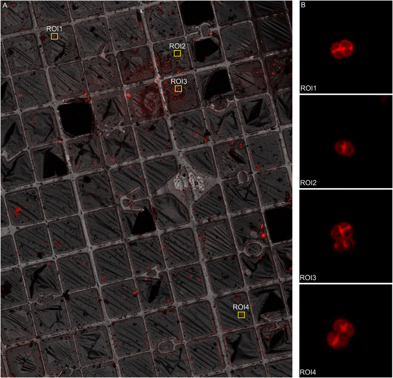
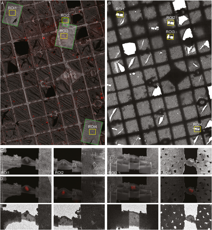
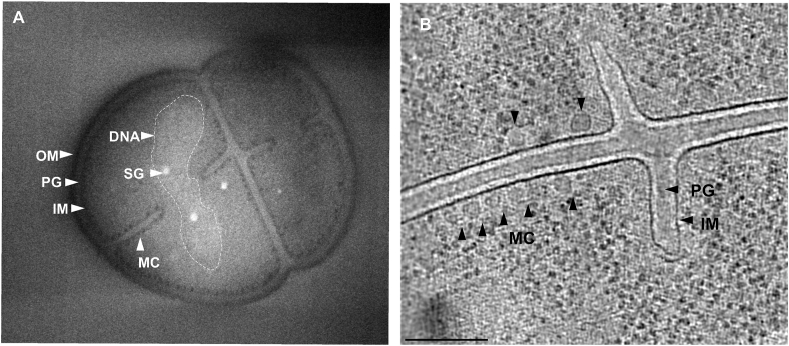
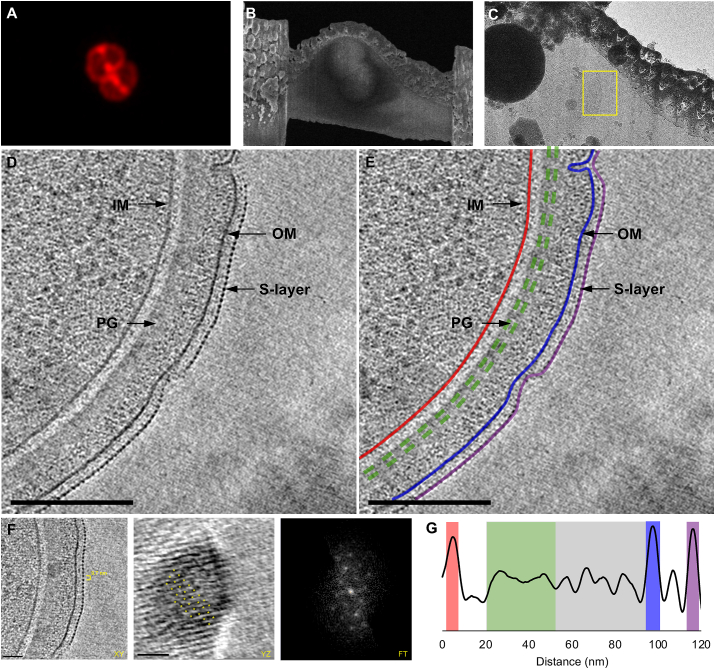
Studying bacterial cell envelope architecture with electron microscopy is challenging due to the poor preservation of microbial ultrastructure with traditional methods. Here, we established and validated a super-resolution cryo-correlative light and electron microscopy (cryo-CLEM) method, and combined it with cryo-focused ion beam (cryo-FIB) milling and scanning electron microscopy (SEM) volume imaging to structurally characterize the bacterium . Subsequent cryo-electron tomography (cryo-ET) revealed an unusual diderm cell envelope architecture with a thick layer of peptidoglycan (PG) between the inner and outer membranes, an additional periplasmic layer, and a proteinaceous surface S-layer. Cells grew in tetrads, and division septa were formed by invagination of the inner membrane (IM), followed by a thick layer of PG. Cytoskeletal filaments, FtsA and FtsZ, were observed at the leading edges of constricting septa. Numerous macromolecular complexes were found associated with the cytoplasmic side of the IM. Altogether, our study revealed several unique ultrastructural features of cells, opening new lines of investigation into the physiology and evolution of the bacterium.

摘要

由于传统方法对微生物超微结构的保存不佳,利用电子显微镜研究细菌细胞包膜结构具有挑战性。在此,我们建立并验证了一种超分辨率冷冻关联光电子显微镜(cryo-CLEM)方法,并将其与冷冻聚焦离子束(cryo-FIB)铣削和扫描电子显微镜(SEM)体积成像相结合,以对该细菌进行结构表征。随后的冷冻电子断层扫描(cryo-ET)揭示了一种不寻常的双膜细胞包膜结构,在内膜和外膜之间有一层厚厚的肽聚糖(PG)、一个额外的周质层和一个蛋白质表面S层。细胞以四联形式生长,分裂隔膜由内膜(IM)内陷形成,随后是一层厚厚的PG。在收缩隔膜的前沿观察到细胞骨架丝、FtsA和FtsZ。发现许多大分子复合物与IM的细胞质侧相关联。总之,我们的研究揭示了该细胞的几个独特超微结构特征,为该细菌的生理学和进化研究开辟了新的途径。

https://cdn.ncbi.nlm.nih.gov/pmc/blobs/7187/8688812/761a2cd93ebc/gr6.jpg
https://cdn.ncbi.nlm.nih.gov/pmc/blobs/7187/8688812/7897a5e48475/ga1.jpg
https://cdn.ncbi.nlm.nih.gov/pmc/blobs/7187/8688812/8b72cec5b081/gr1.jpg
https://cdn.ncbi.nlm.nih.gov/pmc/blobs/7187/8688812/d9ae87ba2aec/gr2.jpg
https://cdn.ncbi.nlm.nih.gov/pmc/blobs/7187/8688812/486a512260fa/gr3.jpg
https://cdn.ncbi.nlm.nih.gov/pmc/blobs/7187/8688812/f3a6361c9cd6/gr4.jpg
https://cdn.ncbi.nlm.nih.gov/pmc/blobs/7187/8688812/451daea0209c/gr5.jpg
https://cdn.ncbi.nlm.nih.gov/pmc/blobs/7187/8688812/761a2cd93ebc/gr6.jpg

相似文献

1
Super-resolution confocal cryo-CLEM with cryo-FIB milling for imaging of .用于……成像的具有低温聚焦离子束铣削的超分辨率共聚焦冷冻扫描透射电子显微镜联用技术
Curr Res Struct Biol. 2021 Dec 13;4:1-9. doi: 10.1016/j.crstbi.2021.12.001. eCollection 2022.
2
Advanced cryo-tomography workflow developments - correlative microscopy, milling automation and cryo-lift-out.先进的冷冻电子断层扫描工作流程发展——相关显微镜技术、铣削自动化和冷冻提取技术。
J Microsc. 2021 Feb;281(2):112-124. doi: 10.1111/jmi.12939. Epub 2020 Jul 2.
3
Cryo-correlative light and electron microscopy workflow for cryo-focused ion beam milled adherent cells.用于冷冻聚焦离子束铣削贴壁细胞的冷冻相关光和电子显微镜工作流程。
Methods Cell Biol. 2021;162:273-302. doi: 10.1016/bs.mcb.2020.12.009. Epub 2021 Feb 17.
4
In situ Microfluidic Cryofixation for Cryo Focused Ion Beam Milling and Cryo Electron Tomography.原位微流控冷冻固定用于冷冻聚焦离子束铣削和冷冻电子断层成像。
Sci Rep. 2019 Dec 13;9(1):19133. doi: 10.1038/s41598-019-55413-2.
5
Reactive oxygen FIB spin milling enables correlative workflow for 3D super-resolution light microscopy and serial FIB/SEM of cultured cells.活性氧纤维铣削实现了用于培养细胞的 3D 超分辨率光显微镜和连续纤维切割/扫描电镜的相关工作流程。
Sci Rep. 2021 Jun 23;11(1):13162. doi: 10.1038/s41598-021-92608-y.
6
A multidomain connector links the outer membrane and cell wall in phylogenetically deep-branching bacteria.一种多结构域连接器连接了系统发育深分枝细菌的外膜和细胞壁。
Proc Natl Acad Sci U S A. 2022 Aug 16;119(33):e2203156119. doi: 10.1073/pnas.2203156119. Epub 2022 Aug 9.
7
Correlative Light and Electron Cryo-Microscopy Workflow Combining Micropatterning, Ice Shield, and an In-Chamber Fluorescence Light Microscope.结合微图案化、冰盾和腔内荧光显微镜的相关光与电子低温显微镜工作流程
Bio Protoc. 2023 Dec 20;13(24):e4901. doi: 10.21769/BioProtoc.4901.
8
Interdigitated immunoglobulin arrays form the hyperstable surface layer of the extremophilic bacterium .指状免疫球蛋白阵列构成了极端嗜热菌超稳定的表面层。
Proc Natl Acad Sci U S A. 2023 Apr 18;120(16):e2215808120. doi: 10.1073/pnas.2215808120. Epub 2023 Apr 12.
9
PIE-scope, integrated cryo-correlative light and FIB/SEM microscopy.PIE-scope,集成冷冻相关的光和 FIB/SEM 显微镜。
Elife. 2019 Jul 1;8:e45919. doi: 10.7554/eLife.45919.
10
Sample Preparation by 3D-Correlative Focused Ion Beam Milling for High-Resolution Cryo-Electron Tomography.通过 3D 相关聚焦离子束铣削进行高分辨率冷冻电子断层成像的样品制备。
J Vis Exp. 2021 Oct 25(176). doi: 10.3791/62886.

引用本文的文献

1
High-throughput cryo-electron tomography enables multiscale visualization of the inner life of microbes.高通量冷冻电子断层扫描技术能够对微生物的内部生命活动进行多尺度可视化。
Curr Opin Struct Biol. 2025 Aug;93:103065. doi: 10.1016/j.sbi.2025.103065. Epub 2025 May 16.
2
A new age in structural S-layer biology: Experimental and in silico milestones.结构表层生物学的新时代:实验与计算机模拟的里程碑。
J Biol Chem. 2025 May 8;301(6):110205. doi: 10.1016/j.jbc.2025.110205.
3
Combining live fluorescence imaging with in situ cryoelectron tomography sheds light on the septation process in .

本文引用的文献

1
CryoSIM: super-resolution 3D structured illumination cryogenic fluorescence microscopy for correlated ultrastructural imaging.CryoSIM:用于相关超微结构成像的超分辨率三维结构光照低温荧光显微镜
Optica. 2020 Jul 13;7(7):802-812. doi: 10.1364/OPTICA.393203. eCollection 2020 Jul 20.
2
Structural analysis of the architecture and in situ localization of the main S-layer complex in Deinococcus radiodurans.结构分析的架构和原位定位的主要 S 层复合物在耐辐射球菌。
Structure. 2021 Nov 4;29(11):1279-1285.e3. doi: 10.1016/j.str.2021.06.014. Epub 2021 Jul 14.
3
Elucidating Protein Translocon Dynamics with Single-Molecule Precision.
将实时荧光成像与原位冷冻电子断层扫描相结合,揭示了……中的隔膜形成过程。
Proc Natl Acad Sci U S A. 2025 May 13;122(19):e2425047122. doi: 10.1073/pnas.2425047122. Epub 2025 May 6.
4
Visualizing the virus world inside the cell by cryo-electron tomography.通过冷冻电子断层扫描技术观察细胞内的病毒世界。
J Virol. 2024 Dec 17;98(12):e0108523. doi: 10.1128/jvi.01085-23. Epub 2024 Nov 4.
5
Recent advances in correlative cryo-light and electron microscopy.关联冷冻光镜和电子显微镜技术的最新进展。
Curr Opin Struct Biol. 2024 Dec;89:102934. doi: 10.1016/j.sbi.2024.102934. Epub 2024 Oct 3.
6
Cell envelope diversity and evolution across the bacterial tree of life.细菌生命之树的细胞包膜多样性和进化。
Nat Microbiol. 2024 Oct;9(10):2475-2487. doi: 10.1038/s41564-024-01812-9. Epub 2024 Sep 18.
7
Liquid Phase Electron Microscopy of Bacterial Ultrastructure.细菌超微结构的液相电子显微镜观察
Small. 2024 Dec;20(50):e2402871. doi: 10.1002/smll.202402871. Epub 2024 Sep 6.
8
Terrabacteria: redefining bacterial envelope diversity, biogenesis and evolution.陆地细菌:重新定义细菌包膜的多样性、生物发生与进化
Nat Rev Microbiol. 2025 Jan;23(1):41-56. doi: 10.1038/s41579-024-01088-0. Epub 2024 Aug 28.
9
CryoET of β-amyloid and tau within postmortem Alzheimer's disease brain.冷冻电镜技术在阿尔茨海默病死后大脑中β-淀粉样蛋白和 tau 的研究。
Nature. 2024 Jul;631(8022):913-919. doi: 10.1038/s41586-024-07680-x. Epub 2024 Jul 10.
10
Structure predictions and functional insights into Amidase_3 domain containing N-acetylmuramyl-L-alanine amidases from Deinococcus indicus DR1.结构预测和功能研究揭示了印度德氏菌 DR1 中酰胺酶_3 结构域含有的 N-乙酰胞壁酰-L-丙氨酸酰胺酶。
BMC Microbiol. 2024 Mar 26;24(1):101. doi: 10.1186/s12866-024-03225-4.
用单分子精度阐明蛋白质易位子动力学。
Trends Cell Biol. 2021 Jul;31(7):569-583. doi: 10.1016/j.tcb.2021.03.009. Epub 2021 Apr 14.
4
Structural, Metabolic and Evolutionary Comparison of Bacterial Endospore and Exospore Formation.细菌内生孢子和外生孢子形成的结构、代谢及进化比较
Front Microbiol. 2021 Mar 9;12:630573. doi: 10.3389/fmicb.2021.630573. eCollection 2021.
5
Functional and pathological amyloid structures in the eyes of 2020 cryo-EM.2020 年冷冻电镜技术下的眼部功能和病理淀粉样结构。
Curr Opin Struct Biol. 2021 Jun;68:184-193. doi: 10.1016/j.sbi.2021.01.006. Epub 2021 Feb 22.
6
Post-correlation on-lamella cryo-CLEM reveals the membrane architecture of lamellar bodies.关联断层成像冷冻电子显微镜技术揭示板层小体的膜结构。
Commun Biol. 2021 Jan 29;4(1):137. doi: 10.1038/s42003-020-01567-z.
7
Unveiling the polarity of actin filaments by cryo-electron tomography.通过冷冻电镜断层成像技术揭示肌动蛋白丝的极性。
Structure. 2021 May 6;29(5):488-498.e4. doi: 10.1016/j.str.2020.12.014. Epub 2021 Jan 20.
8
Structural overview of macromolecular machines involved in ribosome biogenesis.参与核糖体生物发生的大分子机器的结构概述。
Curr Opin Struct Biol. 2021 Apr;67:51-60. doi: 10.1016/j.sbi.2020.09.003. Epub 2020 Oct 21.
9
Serial cryoFIB/SEM Reveals Cytoarchitectural Disruptions in Leigh Syndrome Patient Cells.连续 cryoFIB/SEM 揭示 Leigh 综合征患者细胞的细胞结构紊乱。
Structure. 2021 Jan 7;29(1):82-87.e3. doi: 10.1016/j.str.2020.10.003. Epub 2020 Oct 22.
10
Topaz-Denoise: general deep denoising models for cryoEM and cryoET.Topaz-Denoise:用于 cryoEM 和 cryoET 的通用深度去噪模型。
Nat Commun. 2020 Oct 15;11(1):5208. doi: 10.1038/s41467-020-18952-1.