Dong Daifeng, Ironside Maria, Belleau Emily L, Sun Xiaoqiang, Cheng Chang, Xiong Ge, Nickerson Lisa D, Wang Xiang, Yao Shuqiao, Pizzagalli Diego A
Medical Psychological Center, The Second Xiangya Hospital of Central South University, Changsha, Hunan, P.R. China.
China National Clinical Research Center for Mental Disorders (Xiangya), Changsha, Hunan, P.R. China.
Transl Psychiatry. 2022 Jan 10;12(1):2. doi: 10.1038/s41398-021-01768-y.
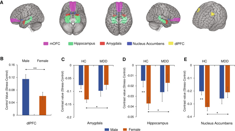
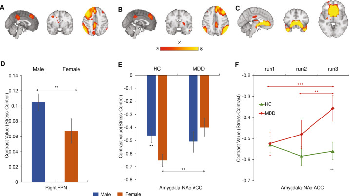
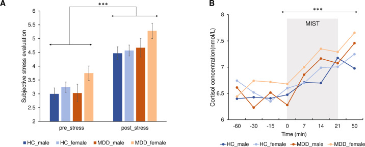
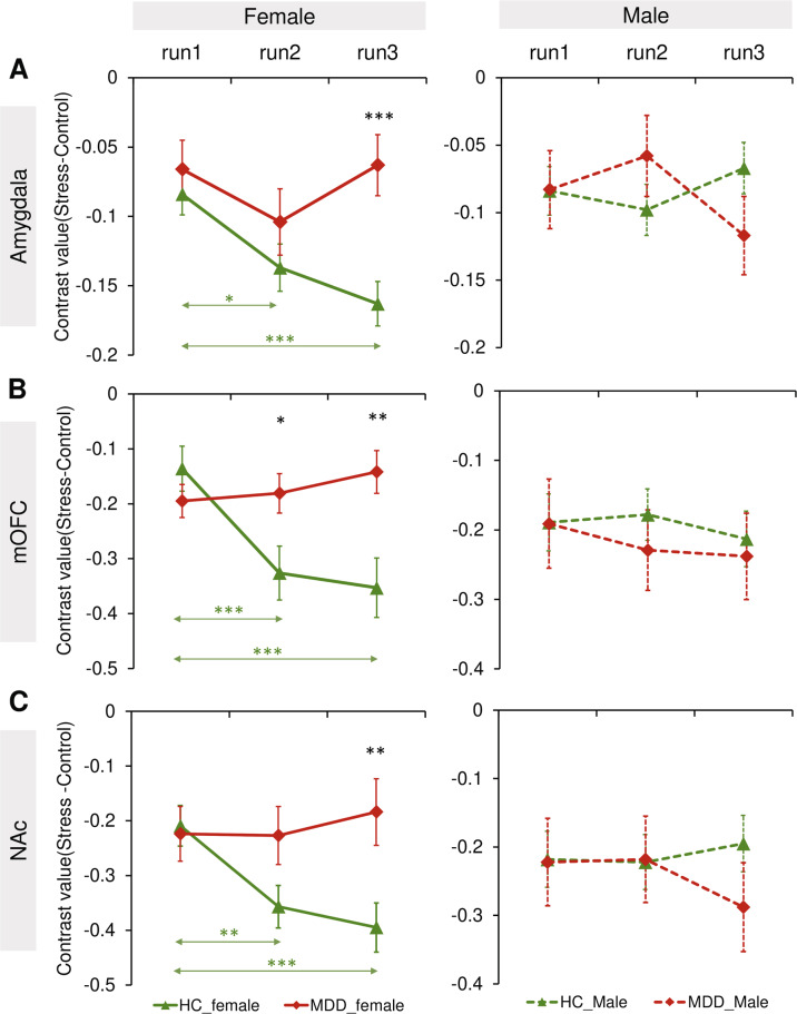
Major Depressive Disorder (MDD) is characterized by increased stress sensitivity. Emerging findings in healthy adults suggest that stress responses within limbic/striatal-prefrontal regions are moderated by sex and unfold over time. Thus, we hypothesized that stress response abnormalities in MDD might be affected by sex and stress exposure time. The Montreal Imaging Stress Task was administered to 124 unmedicated patients with first-episode MDD (76 females) and 243 healthy controls (HC; 137 females) during functional magnetic resonance imaging (fMRI). Based on prior studies, amygdala, hippocampus, medial orbitofrontal cortex (mOFC), nucleus accumbens (NAc) and dorsolateral prefrontal cortex (dlPFC) were selected as a priori regions of interest. In a complementary approach, we probed the effects of stress on the frontoparietal network (FPN) and a network including the amygdala, NAc and anterior cingulate cortex (ACC). Across groups, males exhibited higher dlPFC activity and right FPN amplitude than females. Relative to female HCs, the female MDD group had less deactivation in limbic/striatal regions (amygdala, NAc, hippocampus, Amygdala-NAc-ACC network). Furthermore, unlike female HCs, the female MDD group failed to show a significant increase of deactivation over stress exposure time in the amygdala, mOFC and NAc. Our findings confirm the importance of considering sex differences when investigating neural stress responses. Case-control differences in neural stress responses observed in females (but not males) provide insights into sex differences in the etiology and pathophysiology of depression. The failure to deactivate limbic/NAc regions in depressed females point to dysfunction of adaptive stress responses over stress exposure time.
重度抑郁症(MDD)的特征是应激敏感性增加。健康成年人的新研究结果表明,边缘系统/纹状体-前额叶区域内的应激反应受性别调节,并随时间发展。因此,我们假设MDD中的应激反应异常可能受性别和应激暴露时间的影响。在功能磁共振成像(fMRI)期间,对124名未接受药物治疗的首发MDD患者(76名女性)和243名健康对照者(HC;137名女性)进行了蒙特利尔成像应激任务测试。基于先前的研究,杏仁核、海马体、内侧眶额皮质(mOFC)、伏隔核(NAc)和背外侧前额叶皮质(dlPFC)被选为预先设定的感兴趣区域。采用补充方法,我们探究了应激对额顶叶网络(FPN)以及包括杏仁核、NAc和前扣带回皮质(ACC)的网络的影响。在所有组中,男性的dlPFC活动和右侧FPN振幅均高于女性。相对于女性HC,女性MDD组在边缘系统/纹状体区域(杏仁核、NAc、海马体、杏仁核-NAc-ACC网络)的失活较少。此外,与女性HC不同,女性MDD组在杏仁核、mOFC和NAc中未显示出随着应激暴露时间的延长失活有显著增加。我们的研究结果证实了在研究神经应激反应时考虑性别差异的重要性。在女性(而非男性)中观察到的神经应激反应的病例对照差异为抑郁症的病因和病理生理学中的性别差异提供了见解。抑郁女性边缘系统/NAc区域未能失活表明在应激暴露时间内适应性应激反应功能失调。