Department of Anatomy, School of Basic Medicine, Shanghai University of Traditional Chinese Medicine, Shanghai 201203, China.
Department of Orthopaedics, Shanghai Key Laboratory for Prevention and Treatment of Bone and Joint Diseases, Shanghai Institute of Traumatology and Orthopaedics, Ruijin Hospital, Shanghai Jiao Tong University School of Medicine, Shanghai 200025, China.
Oxid Med Cell Longev. 2022 Jan 29;2022:7530102. doi: 10.1155/2022/7530102. eCollection 2022.
Our study is aimed at investigating the mechanism by which electroacupuncture (EA) promoted nerve regeneration by regulating the release of exosomes and exosome-mediated miRNA-21 (miR-21) transmission. Furthermore, the effects of Schwann cells- (SC-) derived exosomes on the overexpression of miR-21 for the treatment of PNI were investigated.
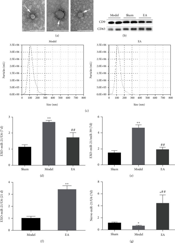
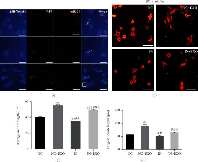
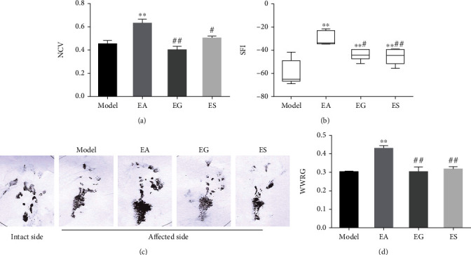
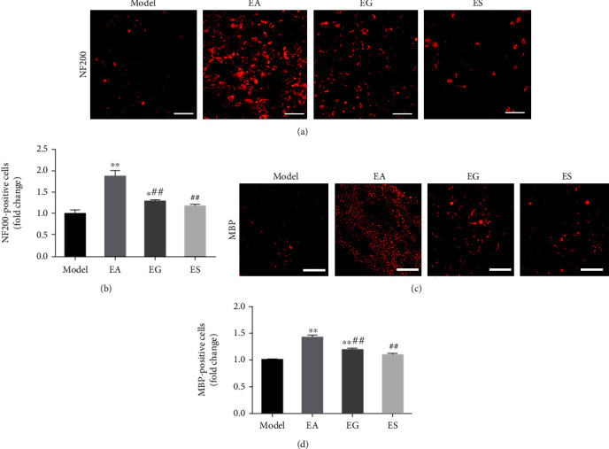
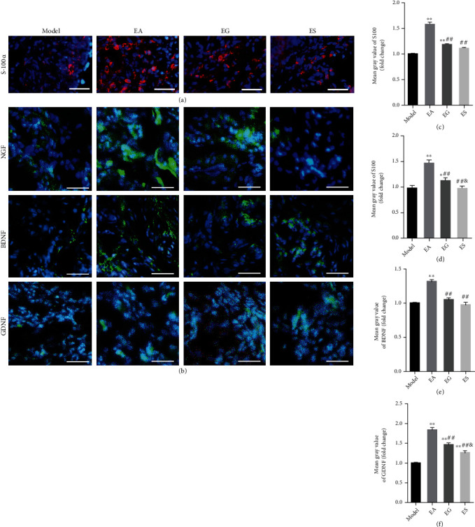
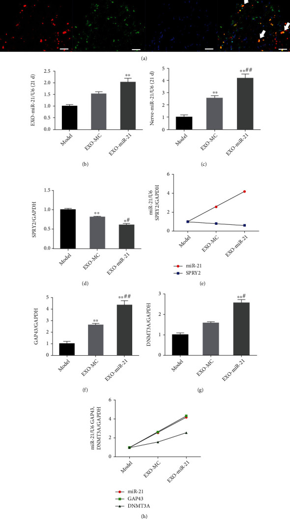
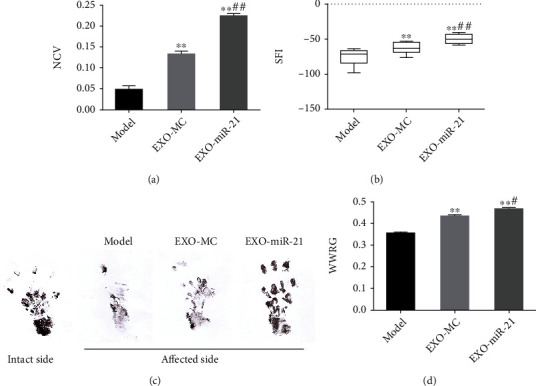
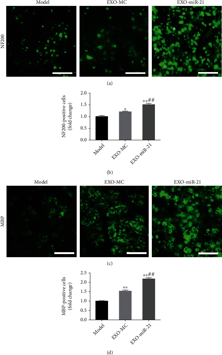
A sciatic nerve injury model of rat was constructed, and the expression of miR-21 in serum exosomes and damaged local nerves was detected using RT-qPCR after EA treatment. The exosomes were identified under a transmission electron microscope and using western blotting analysis. Then, the exosome release inhibitor, GW4869, and the miR-21-5p-sponge used for the knockdown of miR-21 were used to clarify the effects of exosomal miR-21 on nerve regeneration promoted by EA. The nerve conduction velocity recovery rate, sciatic nerve function index, and wet weight ratio of gastrocnemius muscle were determined to evaluate sciatic nerve function recovery. SC proliferation and the level of neurotrophic factors were assessed using immunofluorescence staining, and the expression levels of SPRY2 and miR-21 were detected using RT-qPCR analysis. Subsequently, the transmission of exosomal miR-21 from SC to the axon was verified . Finally, the exosomes derived from the SC infected with the miR-21 overexpression lentivirus were collected and used to treat the rat SNI model to explore the therapeutic role of SC-derived exosomes overexpressing miR-21.
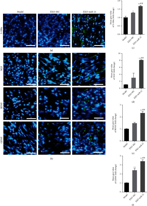
We found that EA inhibited the release of serum exosomal miR-21 in a PNI model of rats during the early stage of PNI, while it promoted its release during later stages. EA enhanced the accumulation of miR-21 in the injured nerve and effectively promoted the recovery of nerve function after PNI. The treatment effect of EA was attenuated when the release of circulating exosomes was inhibited or when miR-21 was downregulated in local injury tissue via the miR-21-5p-sponge. Normal exosomes secreted by SC exhibited the ability to promote the recovery of nerve function, while the overexpression of miR-21 enhanced the effects of the exosomes. In addition, exosomal miR-21 secreted by SC could promote neurite outgrowth .
Our results demonstrated the mechanism of EA on PNI from the perspective of exosome-mediated miR-21 transport and provided a theoretical basis for the use of exosomal miR-21 as a novel strategy for the treatment of PNI.
本研究旨在探讨电针对神经再生的促进作用的机制,即通过调节外泌体的释放及其介导的微小 RNA-21(miR-21)传递。此外,还研究了施万细胞(SCs)衍生的外泌体对 miR-21 过表达的影响,以治疗周围神经损伤(PNI)。
构建大鼠坐骨神经损伤模型,用电针(EA)治疗后,通过 RT-qPCR 检测血清外泌体和损伤局部神经中 miR-21 的表达。在透射电子显微镜下和通过 Western blot 分析鉴定外泌体。然后,使用外泌体释放抑制剂 GW4869 和用于 miR-21 敲低的 miR-21-5p 海绵,以阐明 EA 促进的外泌体 miR-21 对神经再生的影响。通过测定坐骨神经功能指数、神经传导速度恢复率和比目鱼肌湿重比来评估坐骨神经功能的恢复情况。通过免疫荧光染色评估 SC 的增殖和神经营养因子水平,通过 RT-qPCR 分析检测 SPRY2 和 miR-21 的表达水平。随后,验证了外泌体 miR-21 从 SC 向轴突的传递。最后,收集感染 miR-21 过表达慢病毒的 SC 衍生的外泌体,用于治疗大鼠 SNI 模型,以探讨 SC 衍生的过表达 miR-21 的外泌体的治疗作用。
我们发现,在大鼠 PNI 模型中,EA 在 PNI 的早期阶段抑制了血清外泌体 miR-21 的释放,而在后期则促进了其释放。EA 增强了 miR-21 在损伤神经中的积累,并有效促进了 PNI 后神经功能的恢复。当通过 miR-21-5p 海绵抑制循环外泌体的释放或下调局部损伤组织中的 miR-21 时,EA 的治疗效果减弱。SC 分泌的正常外泌体表现出促进神经功能恢复的能力,而过表达 miR-21 增强了外泌体的作用。此外,SC 分泌的外泌体 miR-21 可促进轴突的生长。
本研究从外泌体介导的 miR-21 转运角度探讨了 EA 对 PNI 的作用机制,为将外泌体 miR-21 作为治疗 PNI 的新策略提供了理论依据。