• 文献检索
  • 文档翻译
  • 深度研究
  • 学术资讯
  • Suppr Zotero 插件Zotero 插件
  • 邀请有礼
  • 套餐&价格
  • 历史记录
应用&插件
Suppr Zotero 插件Zotero 插件浏览器插件Mac 客户端Windows 客户端微信小程序
定价
高级版会员购买积分包购买API积分包
服务
文献检索文档翻译深度研究API 文档MCP 服务
关于我们
关于 Suppr公司介绍联系我们用户协议隐私条款
关注我们

Suppr 超能文献

核心技术专利:CN118964589B侵权必究
粤ICP备2023148730 号-1Suppr @ 2026

文献检索

告别复杂PubMed语法,用中文像聊天一样搜索,搜遍4000万医学文献。AI智能推荐,让科研检索更轻松。

立即免费搜索

文件翻译

保留排版,准确专业,支持PDF/Word/PPT等文件格式,支持 12+语言互译。

免费翻译文档

深度研究

AI帮你快速写综述,25分钟生成高质量综述,智能提取关键信息,辅助科研写作。

立即免费体验

通过缺氧施万细胞衍生的外泌体增强周围神经损伤后的神经内再血管化:内皮细胞糖酵解的新见解。

Enhancing intraneural revascularization following peripheral nerve injury through hypoxic Schwann-cell-derived exosomes: an insight into endothelial glycolysis.

机构信息

Department of Neurosurgery, the Third Affiliated Hospital of Sun Yat-sen University, No. 600 Tianhe Road, Guangzhou, Guangdong, 510630, PR China.

Department of Radiology, Guangdong Provincial Hospital of Chinese Medicine, Guangzhou, 510000, China.

出版信息

J Nanobiotechnology. 2024 May 24;22(1):283. doi: 10.1186/s12951-024-02536-y.

DOI:10.1186/s12951-024-02536-y
PMID:38789980
原文链接:https://pmc.ncbi.nlm.nih.gov/articles/PMC11127458/
Abstract

BACKGROUND

Endothelial cell (EC)-driven intraneural revascularization (INRV) and Schwann cells-derived exosomes (SCs-Exos) both play crucial roles in peripheral nerve injury (PNI). However, the interplay between them remains unclear. We aimed to elucidate the effects and underlying mechanisms of SCs-Exos on INRV following PNI.

RESULTS

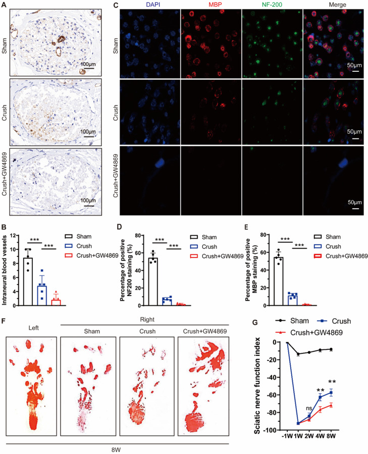
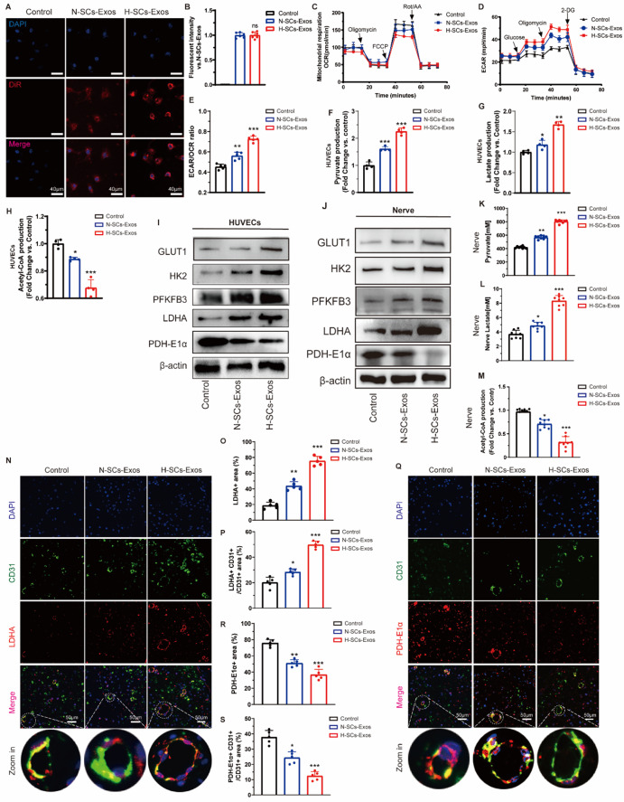
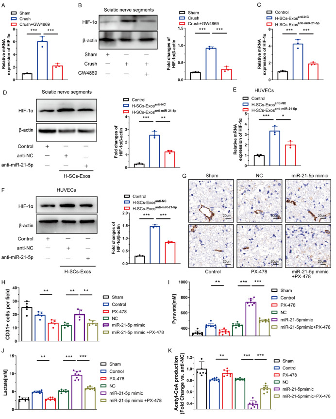
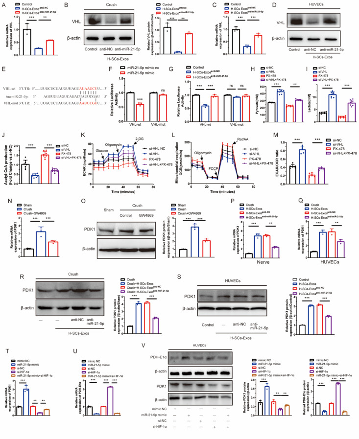
We found that GW4869 inhibited INRV, as well as that normoxic SCs-Exos (N-SCs-Exos) exhibited significant pro-INRV effects in vivo and in vitro that were potentiated by hypoxic SCs-Exos (H-SCs-Exos). Upregulation of glycolysis emerged as a pivotal factor for INRV after PNI, as evidenced by the observation that 3PO administration, a glycolytic inhibitor, inhibited the INRV process in vivo and in vitro. H-SCs-Exos more significantly enhanced extracellular acidification rate/oxygen consumption rate ratio, lactate production, and glycolytic gene expression while simultaneously suppressing acetyl-CoA production and pyruvate dehydrogenase E1 subunit alpha (PDH-E1α) expression than N-SCs-Exos both in vivo and in vitro. Furthermore, we determined that H-SCs-Exos were more enriched with miR-21-5p than N-SCs-Exos. Knockdown of miR-21-5p significantly attenuated the pro-glycolysis and pro-INRV effects of H-SCs-Exos. Mechanistically, miR-21-5p orchestrated EC metabolism in favor of glycolysis by targeting von Hippel-Lindau/hypoxia-inducible factor-1α and PDH-E1α, thereby enhancing hypoxia-inducible factor-1α-mediated glycolysis and inhibiting PDH-E1α-mediated oxidative phosphorylation.

CONCLUSION

This study unveiled a novel intrinsic mechanism of pro-INRV after PNI, providing a promising therapeutic target for post-injury peripheral nerve regeneration and repair.

摘要

背景

内皮细胞(EC)驱动的神经内再血管化(INRV)和施万细胞衍生的外泌体(SCs-Exos)在外周神经损伤(PNI)中都起着至关重要的作用。然而,它们之间的相互作用尚不清楚。我们旨在阐明SCs-Exos 对 PNI 后 INRV 的影响及其潜在机制。

结果

我们发现 GW4869 抑制 INRV,并且在体内和体外,缺氧SCs-Exos(H-SCs-Exos)增强了正常氧SCs-Exos(N-SCs-Exos)的显著促 INRV 作用。PNI 后糖酵解的上调成为 INRV 的关键因素,因为观察到糖酵解抑制剂 3PO 给药在体内和体外抑制 INRV 过程。H-SCs-Exos 比 N-SCs-Exos 更显著地增强细胞外酸化率/耗氧量比、乳酸产生和糖酵解基因表达,同时抑制乙酰辅酶 A 产生和丙酮酸脱氢酶 E1 亚基 alpha(PDH-E1α)表达,无论是在体内还是体外。此外,我们确定 H-SCs-Exos 比 N-SCs-Exos 更富含 miR-21-5p。miR-21-5p 的敲低显著减弱了 H-SCs-Exos 的促糖酵解和促 INRV 作用。在机制上,miR-21-5p 通过靶向 von Hippel-Lindau/缺氧诱导因子-1α和 PDH-E1α 来协调 EC 代谢有利于糖酵解,从而增强缺氧诱导因子-1α 介导的糖酵解和抑制 PDH-E1α 介导的氧化磷酸化。

结论

这项研究揭示了 PNI 后促 INRV 的一种新的内在机制,为损伤后周围神经再生和修复提供了有希望的治疗靶点。

https://cdn.ncbi.nlm.nih.gov/pmc/blobs/2c1f/11127458/056cfbff224e/12951_2024_2536_Fig8_HTML.jpg
https://cdn.ncbi.nlm.nih.gov/pmc/blobs/2c1f/11127458/55c2d5cc06e5/12951_2024_2536_Fig1_HTML.jpg
https://cdn.ncbi.nlm.nih.gov/pmc/blobs/2c1f/11127458/3ae663426bfd/12951_2024_2536_Fig2_HTML.jpg
https://cdn.ncbi.nlm.nih.gov/pmc/blobs/2c1f/11127458/87e82cd910a9/12951_2024_2536_Fig3_HTML.jpg
https://cdn.ncbi.nlm.nih.gov/pmc/blobs/2c1f/11127458/c0436b15d2e7/12951_2024_2536_Fig4_HTML.jpg
https://cdn.ncbi.nlm.nih.gov/pmc/blobs/2c1f/11127458/33272429a887/12951_2024_2536_Fig5_HTML.jpg
https://cdn.ncbi.nlm.nih.gov/pmc/blobs/2c1f/11127458/3648e3fc59b6/12951_2024_2536_Fig6_HTML.jpg
https://cdn.ncbi.nlm.nih.gov/pmc/blobs/2c1f/11127458/81fea39a0c86/12951_2024_2536_Fig7_HTML.jpg
https://cdn.ncbi.nlm.nih.gov/pmc/blobs/2c1f/11127458/056cfbff224e/12951_2024_2536_Fig8_HTML.jpg

相似文献

1
Enhancing intraneural revascularization following peripheral nerve injury through hypoxic Schwann-cell-derived exosomes: an insight into endothelial glycolysis.通过缺氧施万细胞衍生的外泌体增强周围神经损伤后的神经内再血管化:内皮细胞糖酵解的新见解。
J Nanobiotechnology. 2024 May 24;22(1):283. doi: 10.1186/s12951-024-02536-y.
2
Study on the Role and Mechanism of Exosomes Derived from Dental Pulp Stem Cells in Promoting Regeneration of Myelin Sheath in Rats with Sciatic Nerve Injury.牙髓干细胞来源的外泌体在促进坐骨神经损伤大鼠髓鞘再生中的作用及机制研究。
Mol Neurobiol. 2024 Sep;61(9):6175-6188. doi: 10.1007/s12035-024-03960-9. Epub 2024 Jan 29.
3
Down-regulation miR-146a-5p in Schwann cell-derived exosomes induced macrophage M1 polarization by impairing the inhibition on TRAF6/NF-κB pathway after peripheral nerve injury.周围神经损伤后,雪旺细胞衍生外泌体中miR-146a-5p的下调通过削弱对TRAF6/NF-κB通路的抑制作用诱导巨噬细胞M1极化。
Exp Neurol. 2023 Apr;362:114295. doi: 10.1016/j.expneurol.2022.114295. Epub 2022 Dec 6.
4
Exosomes produced by adipose-derived stem cells inhibit schwann cells autophagy and promote the regeneration of the myelin sheath.脂肪来源干细胞分泌的外泌体抑制许旺细胞自噬,促进髓鞘再生。
Int J Biochem Cell Biol. 2021 Mar;132:105921. doi: 10.1016/j.biocel.2021.105921. Epub 2021 Jan 6.
5
Exosome-Mediated miR-21 Was Involved in the Promotion of Structural and Functional Recovery Effect Produced by Electroacupuncture in Sciatic Nerve Injury.外泌体介导的 miR-21 参与电针对坐骨神经损伤的结构和功能恢复作用的促进。
Oxid Med Cell Longev. 2022 Jan 29;2022:7530102. doi: 10.1155/2022/7530102. eCollection 2022.
6
Hypoxic Bone Mesenchymal Stem Cell-Derived Exosomes Direct Schwann Cells Proliferation, Migration, and Paracrine to Accelerate Facial Nerve Regeneration via circRNA_Nkd2/miR-214-3p/MED19 Axis.缺氧骨髓间充质干细胞衍生的外泌体通过 circRNA_Nkd2/miR-214-3p/MED19 轴直接诱导许旺细胞增殖、迁移和旁分泌,从而加速面神经再生。
Int J Nanomedicine. 2024 Feb 12;19:1409-1429. doi: 10.2147/IJN.S443036. eCollection 2024.
7
OTULIN of exosomes derived from Schwann cells promotes peripheral nerve injury repair by regulating macrophage polarization via deubiquitination of ERBB2.施万细胞来源的外泌体 OTULIN 通过去泛素化 ERBB2 调节巨噬细胞极化促进周围神经损伤修复。
Neurosci Lett. 2024 Jun 11;833:137813. doi: 10.1016/j.neulet.2024.137813. Epub 2024 May 7.
8
Endothelial cell-derived exosomes boost and maintain repair-related phenotypes of Schwann cells via miR199-5p to promote nerve regeneration.内皮细胞衍生的外泌体通过 miR199-5p 促进和维持施万细胞的修复相关表型,从而促进神经再生。
J Nanobiotechnology. 2023 Jan 9;21(1):10. doi: 10.1186/s12951-023-01767-9.
9
The effects of exosomes originating from different cell sources on the differentiation of bone marrow mesenchymal stem cells into schwann cells.源自不同细胞来源的外泌体对骨髓间充质干细胞向施万细胞分化的影响。
J Nanobiotechnology. 2024 May 3;22(1):220. doi: 10.1186/s12951-024-02450-3.
10
Therapeutic effect of exosomes derived from Schwann cells in the repair of peripheral nerve injury.施万细胞来源的外泌体在周围神经损伤修复中的治疗效果。
Life Sci. 2024 Nov 15;357:123086. doi: 10.1016/j.lfs.2024.123086. Epub 2024 Sep 30.

引用本文的文献

1
The Role of Vascularization in Nerve Regeneration: Mechanistic and Therapeutic Perspectives.血管化在神经再生中的作用:机制与治疗前景
Int J Mol Sci. 2025 Aug 29;26(17):8395. doi: 10.3390/ijms26178395.
2
Pathologic and Therapeutic Schwann Cells.病理性与治疗性施万细胞
Cells. 2025 Aug 28;14(17):1336. doi: 10.3390/cells14171336.
3
Extracellular vesicles derived from Schwann cells to enhance bone and dental tissue regeneration: a literature review.源自雪旺细胞的细胞外囊泡促进骨和牙组织再生:文献综述

本文引用的文献

1
Neural tissue-engineered prevascularization enhances peripheral neuroregeneration via rapid vascular inosculation.神经组织工程预血管化通过快速血管吻合促进周围神经再生。
Mater Today Bio. 2023 Jun 30;21:100718. doi: 10.1016/j.mtbio.2023.100718. eCollection 2023 Aug.
2
AP2α negatively regulates PDHA1 in cervical cancer cells to promote aggressive features and aerobic glycolysis in vitro and in vivo.AP2α 在宫颈癌细胞中负调控 PDHA1,以促进体外和体内侵袭性特征和有氧糖酵解。
J Gynecol Oncol. 2023 Sep;34(5):e59. doi: 10.3802/jgo.2023.34.e59. Epub 2023 Apr 5.
3
Skin precursor-derived Schwann cells accelerate in vivo prevascularization of tissue-engineered nerves to promote peripheral nerve regeneration.
J Nanobiotechnology. 2025 Jul 11;23(1):502. doi: 10.1186/s12951-025-03585-7.
4
The Application of Stem Cells and Exosomes in Promoting Nerve Conduits for Peripheral Nerve Repair.干细胞和外泌体在促进周围神经修复神经导管中的应用
Biomater Res. 2025 Apr 14;29:0160. doi: 10.34133/bmr.0160. eCollection 2025.
5
Strategies for promoting neurovascularization in bone regeneration.促进骨再生中神经血管化的策略。
Mil Med Res. 2025 Mar 3;12(1):9. doi: 10.1186/s40779-025-00596-1.
6
Molecular and cellular mechanisms underlying peripheral nerve injury-induced cellular ecological shifts: Implications for neuroregeneration.外周神经损伤诱导细胞生态转变的分子和细胞机制:对神经再生的启示
IBRO Neurosci Rep. 2024 Dec 28;18:120-129. doi: 10.1016/j.ibneur.2024.12.013. eCollection 2025 Jun.
7
ATP5J regulates microglial activation via mitochondrial dysfunction, exacerbating neuroinflammation in intracerebral hemorrhage.ATP5J通过线粒体功能障碍调节小胶质细胞活化,加剧脑出血中的神经炎症。
Front Immunol. 2024 Dec 13;15:1509370. doi: 10.3389/fimmu.2024.1509370. eCollection 2024.
皮肤前体细胞来源的雪旺细胞可加速组织工程神经的体内血管形成前过程,以促进周围神经再生。
Glia. 2023 Jul;71(7):1755-1769. doi: 10.1002/glia.24367. Epub 2023 Mar 27.
4
Overactivation of Norepinephrine-β2-Adrenergic Receptor Axis Promotes Corneal Neovascularization.去甲肾上腺素-β2-肾上腺素能受体轴的过度激活促进角膜新生血管形成。
Invest Ophthalmol Vis Sci. 2023 Mar 1;64(3):20. doi: 10.1167/iovs.64.3.20.
5
S-Propargyl-Cysteine Ameliorates Peripheral Nerve Injury through Microvascular Reconstruction.S-炔丙基-半胱氨酸通过微血管重建改善周围神经损伤。
Antioxidants (Basel). 2023 Jan 28;12(2):294. doi: 10.3390/antiox12020294.
6
Enhancement of glycolysis-dependent DNA repair regulated by FOXO1 knockdown via PFKFB3 attenuates hyperglycemia-induced endothelial oxidative stress injury.敲低 FOXO1 增强糖酵解依赖性 DNA 修复并通过 PFKFB3 减轻高糖诱导的内皮氧化应激损伤。
Redox Biol. 2023 Feb;59:102589. doi: 10.1016/j.redox.2022.102589. Epub 2022 Dec 25.
7
A multifunctional neuromodulation platform utilizing Schwann cell-derived exosomes orchestrates bone microenvironment via immunomodulation, angiogenesis and osteogenesis.一种利用雪旺细胞衍生外泌体的多功能神经调节平台通过免疫调节、血管生成和成骨作用来协调骨微环境。
Bioact Mater. 2022 Nov 14;23:206-222. doi: 10.1016/j.bioactmat.2022.10.018. eCollection 2023 May.
8
Small extracellular vesicles derived from human adipose-derived stem cells regulate energetic metabolism through the activation of YAP/TAZ pathway facilitating angiogenesis.源自人脂肪干细胞的小细胞外囊泡通过激活YAP/TAZ途径促进血管生成,从而调节能量代谢。
Cell Biol Int. 2023 Feb;47(2):451-466. doi: 10.1002/cbin.11938. Epub 2022 Oct 23.
9
The hypoxia response pathway promotes PEP carboxykinase and gluconeogenesis in C. elegans.缺氧反应通路促进秀丽隐杆线虫中的 PEP 羧激酶和糖异生。
Nat Commun. 2022 Oct 18;13(1):6168. doi: 10.1038/s41467-022-33849-x.
10
The Emerging Role of Pericyte-Derived Extracellular Vesicles in Vascular and Neurological Health.周细胞衍生细胞外囊泡在血管和神经健康中的新作用。
Cells. 2022 Oct 2;11(19):3108. doi: 10.3390/cells11193108.