Department of Hepatobiliary, Pancreatic, and Splenic Surgery, Hebei General Hospital, No. 348 Heping West Road, Shijiazhuang, 050000, China.
World J Surg Oncol. 2022 Feb 27;20(1):55. doi: 10.1186/s12957-022-02525-1.
Primary undifferentiated pleomorphic sarcoma (UPS) of the pancreas is an exceedingly rare malignant tumor, with only 15 cases have been reported in the medical literature. At present, clinicians have poor recognition of the tumor, the epidemiology, diagnosis, and treatment of this disease have yet not been established.
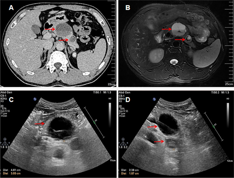
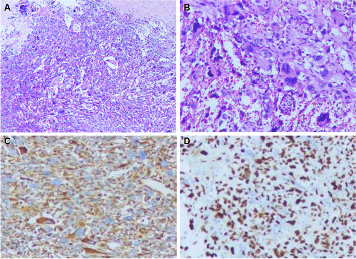
In this report, we depict the clinical and imaging characteristics of a 37-year-old man presenting with a primarily cystic UPS. The patient complained of epigastric pain and distention over 20 days. Abdominal CT and pancreatic magnetic resonance imaging revealed cystic and cystic solid masses in the pancreatic body and tail. An abdominal ultrasound echogram revealed the mass in the body of the pancreas to be cystic with separation echo inside, and the wall was thick, not smooth. Besides, a hypoechoic mass was seen in the tail area of the pancreas with an inhomogeneous echoic pattern, containing small patches of no echo zone in the central. Microscopically, spindle fibroblast-like cells are arranged in a characteristic storiform pattern with pleomorphic and multinucleated cells. Immunohistochemically, tumor cells were positive for CD68 and vimentin. Seven months postoperatively, he was diagnosed with pulmonary lymph node metastasis and died 5 months later. Combined with this case report, we also reviewed the literature regarding UPS of the pancreas.
As we know, this is the first report on ultrasonography findings of pancreatic UPS. Despite there are no distinctive manifestation of UPS, a solid cystic lesion on ultrasonography or a hypodense area in the lesion on T2-weighted imaging, should be considered for differential diagnosis with pancreatic UPS. We believe this article may add some ideas into the diagnosis and therapy of patients with this tumor.
胰腺原发性未分化多形性肉瘤(UPS)是一种极为罕见的恶性肿瘤,文献中仅报道了 15 例。目前,临床医生对该肿瘤的认识较差,尚未建立该疾病的流行病学、诊断和治疗方法。
本报告描述了一名 37 岁男性的临床和影像学特征,该患者表现为主要囊性 UPS。患者诉上腹疼痛和膨胀超过 20 天。腹部 CT 和胰腺磁共振成像显示胰腺体尾部囊性和囊实性肿块。腹部超声回声显示胰腺体部肿块为囊性,内部有分隔回声,壁不光滑。此外,胰腺尾部可见低回声肿块,回声不均匀,中央可见小片状无回声区。显微镜下,梭形成纤维细胞样细胞呈特征性的交织状排列,有异型性和多核细胞。免疫组化染色显示肿瘤细胞 CD68 和波形蛋白阳性。术后 7 个月,患者被诊断为肺淋巴结转移,5 个月后死亡。结合本病例报告,我们还复习了有关胰腺 UPS 的文献。
据我们所知,这是首例关于胰腺 UPS 超声表现的报告。尽管 UPS 没有明显的表现,但超声检查显示为实性囊性病变,或 T2 加权成像显示病变为低信号区,应考虑与胰腺 UPS 进行鉴别诊断。我们相信这篇文章可能会为该肿瘤的诊断和治疗提供一些思路。