Xin Dan-Qing, Zhao Yi-Jing, Li Ting-Ting, Ke Hong-Fei, Gai Cheng-Cheng, Guo Xiao-Fan, Chen Wen-Qiang, Liu De-Xiang, Wang Zhen
Department of Physiology, School of Basic Medical Sciences, Cheeloo College of Medicine, Shandong University, Jinan, Shandong Province, China.
Department of Neurology, Loma Linda University Health, Loma Linda, CA, USA.
Neural Regen Res. 2022 Oct;17(10):2238-2246. doi: 10.4103/1673-5374.336871.
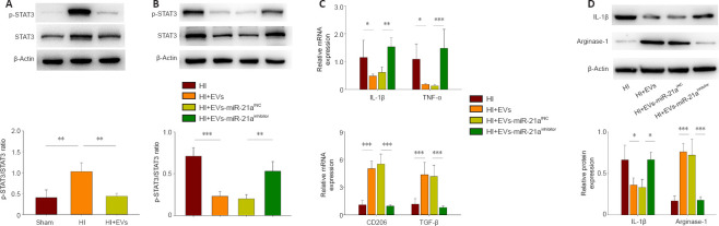
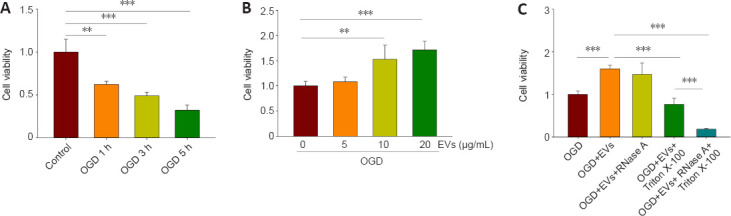
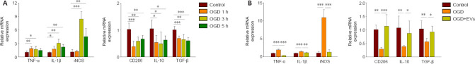
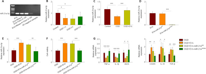
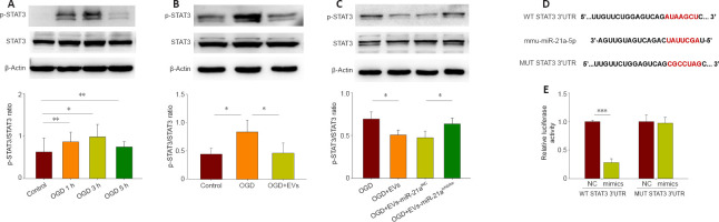
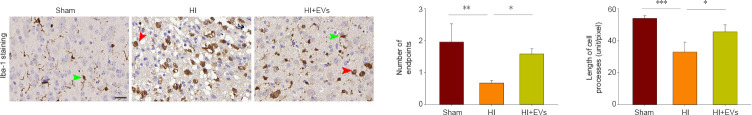
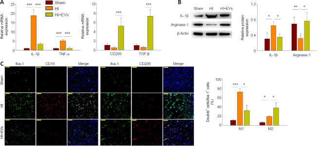
Extracellular vesicles (EVs) from mesenchymal stromal cells (MSCs) have previously been shown to protect against brain injury caused by hypoxia-ischemia (HI). The neuroprotective effects have been found to relate to the anti-inflammatory effects of EVs. However, the underlying mechanisms have not previously been determined. In this study, we induced oxygen-glucose deprivation in BV-2 cells (a microglia cell line), which mimics HI in vitro, and found that treatment with MSCs-EVs increased the cell viability. The treatment was also found to reduce the expression of pro-inflammatory cytokines, induce the polarization of microglia towards the M2 phenotype, and suppress the phosphorylation of selective signal transducer and activator of transcription 3 (STAT3) in the microglia. These results were also obtained in vivo using neonatal mice with induced HI. We investigated the potential role of miR-21a-5p in mediating these effects, as it is the most highly expressed miRNA in MSCs-EVs and interacts with the STAT3 pathway. We found that treatment with MSCs-EVs increased the levels of miR-21a-5p in BV-2 cells, which had been lowered following oxygen-glucose deprivation. When the level of miR-21a-5p in the MSCs-EVs was reduced, the effects on microglial polarization and STAT3 phosphorylation were reduced, for both the in vitro and in vivo HI models. These results indicate that MSCs-EVs attenuate HI brain injury in neonatal mice by shuttling miR-21a-5p, which induces microglial M2 polarization by targeting STAT3.
间充质基质细胞(MSC)来源的细胞外囊泡(EV)此前已被证明可预防缺氧缺血(HI)所致的脑损伤。已发现其神经保护作用与EV的抗炎作用有关。然而,其潜在机制此前尚未确定。在本研究中,我们在BV-2细胞(一种小胶质细胞系)中诱导氧糖剥夺,该过程在体外模拟HI,发现用MSC-EV处理可提高细胞活力。还发现该处理可降低促炎细胞因子的表达,诱导小胶质细胞向M2表型极化,并抑制小胶质细胞中选择性信号转导和转录激活因子3(STAT3)的磷酸化。在体内使用诱导HI的新生小鼠也获得了这些结果。我们研究了miR-21a-5p在介导这些效应中的潜在作用,因为它是MSC-EV中表达最高的miRNA且与STAT3通路相互作用。我们发现用MSC-EV处理可提高氧糖剥夺后BV-2细胞中miR-21a-5p的水平。当降低MSC-EV中miR-21a-5p的水平时,对于体外和体内HI模型,对小胶质细胞极化和STAT3磷酸化的影响均降低。这些结果表明,MSC-EV通过转运miR-21a-5p减轻新生小鼠的HI脑损伤,miR-21a-5p通过靶向STAT3诱导小胶质细胞M2极化。