• 文献检索
  • 文档翻译
  • 深度研究
  • 学术资讯
  • Suppr Zotero 插件Zotero 插件
  • 邀请有礼
  • 套餐&价格
  • 历史记录
应用&插件
Suppr Zotero 插件Zotero 插件浏览器插件Mac 客户端Windows 客户端微信小程序
定价
高级版会员购买积分包购买API积分包
服务
文献检索文档翻译深度研究API 文档MCP 服务
关于我们
关于 Suppr公司介绍联系我们用户协议隐私条款
关注我们

Suppr 超能文献

核心技术专利:CN118964589B侵权必究
粤ICP备2023148730 号-1Suppr @ 2026

文献检索

告别复杂PubMed语法,用中文像聊天一样搜索,搜遍4000万医学文献。AI智能推荐,让科研检索更轻松。

立即免费搜索

文件翻译

保留排版,准确专业,支持PDF/Word/PPT等文件格式,支持 12+语言互译。

免费翻译文档

深度研究

AI帮你快速写综述,25分钟生成高质量综述,智能提取关键信息,辅助科研写作。

立即免费体验

基于深度学习的 CT 胰腺自动分割与体积测量。

Automated pancreas segmentation and volumetry using deep neural network on computed tomography.

机构信息

Department of Health Sciences and Technology, Gachon Advanced Institute for Health Sciences and Technology (GAIHST), Gachon University, 1342, Seongnamdaero, Sujeong-gu, Seongnam-si, 13120, Republic of Korea.

Department of Biomedical Engineering, Gil Medical Center, Gachon University College of Medicine, 38-13, Dokjeon-ro 3beon-gil, Namdong-gu, Incheon, 21565, Republic of Korea.

出版信息

Sci Rep. 2022 Mar 8;12(1):4075. doi: 10.1038/s41598-022-07848-3.

DOI:10.1038/s41598-022-07848-3
PMID:35260710
原文链接:https://pmc.ncbi.nlm.nih.gov/articles/PMC8904764/
Abstract

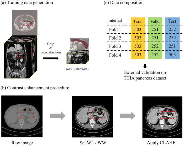
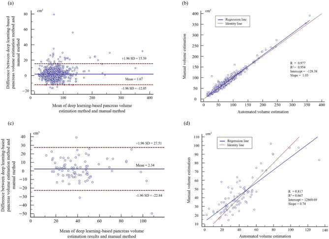
Pancreas segmentation is necessary for observing lesions, analyzing anatomical structures, and predicting patient prognosis. Therefore, various studies have designed segmentation models based on convolutional neural networks for pancreas segmentation. However, the deep learning approach is limited by a lack of data, and studies conducted on a large computed tomography dataset are scarce. Therefore, this study aims to perform deep-learning-based semantic segmentation on 1006 participants and evaluate the automatic segmentation performance of the pancreas via four individual three-dimensional segmentation networks. In this study, we performed internal validation with 1,006 patients and external validation using the cancer imaging archive pancreas dataset. We obtained mean precision, recall, and dice similarity coefficients of 0.869, 0.842, and 0.842, respectively, for internal validation via a relevant approach among the four deep learning networks. Using the external dataset, the deep learning network achieved mean precision, recall, and dice similarity coefficients of 0.779, 0.749, and 0.735, respectively. We expect that generalized deep-learning-based systems can assist clinical decisions by providing accurate pancreatic segmentation and quantitative information of the pancreas for abdominal computed tomography.

摘要

胰腺分割对于观察病变、分析解剖结构和预测患者预后是必要的。因此,许多研究都基于卷积神经网络设计了胰腺分割模型。然而,深度学习方法受到数据缺乏的限制,而且在大型 CT 数据集上进行的研究很少。因此,本研究旨在对 1006 名参与者进行基于深度学习的语义分割,并通过四个独立的三维分割网络评估胰腺的自动分割性能。在本研究中,我们使用 1006 名患者进行内部验证,并使用癌症成像档案胰腺数据集进行外部验证。我们通过四个深度学习网络中的相关方法获得了内部验证的平均精度、召回率和 Dice 相似系数分别为 0.869、0.842 和 0.842。使用外部数据集,深度学习网络的平均精度、召回率和 Dice 相似系数分别为 0.779、0.749 和 0.735。我们期望基于广义深度学习的系统能够通过提供腹部 CT 中胰腺的准确分割和定量信息来辅助临床决策。

https://cdn.ncbi.nlm.nih.gov/pmc/blobs/de6b/8904764/a0db86fe572f/41598_2022_7848_Fig4_HTML.jpg
https://cdn.ncbi.nlm.nih.gov/pmc/blobs/de6b/8904764/64b726d7e6e0/41598_2022_7848_Fig1_HTML.jpg
https://cdn.ncbi.nlm.nih.gov/pmc/blobs/de6b/8904764/8322f8af4f97/41598_2022_7848_Fig2_HTML.jpg
https://cdn.ncbi.nlm.nih.gov/pmc/blobs/de6b/8904764/954879bac090/41598_2022_7848_Fig3_HTML.jpg
https://cdn.ncbi.nlm.nih.gov/pmc/blobs/de6b/8904764/a0db86fe572f/41598_2022_7848_Fig4_HTML.jpg

相似文献

1
Automated pancreas segmentation and volumetry using deep neural network on computed tomography.基于深度学习的 CT 胰腺自动分割与体积测量。
Sci Rep. 2022 Mar 8;12(1):4075. doi: 10.1038/s41598-022-07848-3.
2
Spatial aggregation of holistically-nested convolutional neural networks for automated pancreas localization and segmentation.整体嵌套卷积神经网络的空间聚合用于自动胰腺定位和分割。
Med Image Anal. 2018 Apr;45:94-107. doi: 10.1016/j.media.2018.01.006. Epub 2018 Feb 1.
3
Two-stage deep learning model for fully automated pancreas segmentation on computed tomography: Comparison with intra-reader and inter-reader reliability at full and reduced radiation dose on an external dataset.基于 CT 的全自动胰腺分割的两阶段深度学习模型:在外部数据集上比较全剂量和低剂量下的同读者和异读者可靠性。
Med Phys. 2021 May;48(5):2468-2481. doi: 10.1002/mp.14782. Epub 2021 Mar 16.
4
Technical and Clinical Factors Affecting Success Rate of a Deep Learning Method for Pancreas Segmentation on CT.技术和临床因素对 CT 胰腺分割深度学习方法成功率的影响。
Acad Radiol. 2020 May;27(5):689-695. doi: 10.1016/j.acra.2019.08.014. Epub 2019 Sep 16.
5
nnU-Net-Based Pancreas Segmentation and Volume Measurement on CT Imaging in Patients with Pancreatic Cancer.基于 nnU-Net 的胰腺癌 CT 图像胰腺分割与体积测量。
Acad Radiol. 2024 Jul;31(7):2784-2794. doi: 10.1016/j.acra.2024.01.004. Epub 2024 Feb 12.
6
Extension-contraction transformation network for pancreas segmentation in abdominal CT scans.用于腹部CT扫描中胰腺分割的伸缩变换网络
Comput Biol Med. 2023 Jan;152:106410. doi: 10.1016/j.compbiomed.2022.106410. Epub 2022 Dec 5.
7
Pancreas Segmentation in Abdominal CT Scans using Inter-/Intra-Slice Contextual Information with a Cascade Neural Network.利用层间/层内上下文信息和级联神经网络进行腹部CT扫描中的胰腺分割
Annu Int Conf IEEE Eng Med Biol Soc. 2019 Jul;2019:5937-5940. doi: 10.1109/EMBC.2019.8856774.
8
Improving the slice interaction of 2.5D CNN for automatic pancreas segmentation.改进用于自动胰腺分割的2.5D卷积神经网络的切片交互。
Med Phys. 2020 Nov;47(11):5543-5554. doi: 10.1002/mp.14303. Epub 2020 Oct 15.
9
A Two-Phase Approach using Mask R-CNN and 3D U-Net for High-Accuracy Automatic Segmentation of Pancreas in CT Imaging.基于 Mask R-CNN 和 3D U-Net 的双阶段方法,实现 CT 成像中胰腺的高精度自动分割。
Comput Methods Programs Biomed. 2021 Aug;207:106141. doi: 10.1016/j.cmpb.2021.106141. Epub 2021 May 5.
10
Development and Validation of a Deep Learning System for Segmentation of Abdominal Muscle and Fat on Computed Tomography.基于深度学习的 CT 图像腹部肌肉和脂肪分割系统的建立与验证
Korean J Radiol. 2020 Jan;21(1):88-100. doi: 10.3348/kjr.2019.0470.

引用本文的文献

1
Progress in fully automated abdominal CT interpretation-an update over the past decade.全自动化腹部CT解读的进展——过去十年的最新情况
Abdom Radiol (NY). 2025 Jul 8. doi: 10.1007/s00261-025-05094-5.
2
Automated pancreatic extracellular volume fraction as a biomarker for microangiopathy in type 2 diabetes mellitus.自动化胰腺细胞外容积分数作为2型糖尿病微血管病变的生物标志物
BMC Med Imaging. 2025 Jul 1;25(1):226. doi: 10.1186/s12880-025-01731-z.
3
Artificial Intelligence-Augmented Imaging for Early Pancreatic Cancer Detection.用于早期胰腺癌检测的人工智能增强成像

本文引用的文献

1
Reproducibility of automated habenula segmentation via deep learning in major depressive disorder and normal controls with 7 Tesla MRI.7T MRI 下深度学习在重度抑郁症和正常对照者中海马结构自动分割的可重复性。
Sci Rep. 2021 Jun 29;11(1):13445. doi: 10.1038/s41598-021-92952-z.
2
Multi-scale U-like network with attention mechanism for automatic pancreas segmentation.基于注意力机制的多尺度 U 型网络用于胰腺自动分割。
PLoS One. 2021 May 27;16(5):e0252287. doi: 10.1371/journal.pone.0252287. eCollection 2021.
3
A review of deep learning based methods for medical image multi-organ segmentation.
Visc Med. 2025 May 28:1-9. doi: 10.1159/000546603.
4
The Laryngovibrogram as a normalized spatiotemporal representation of vocal fold dynamics.喉振动图作为声带动力学的标准化时空表征。
Sci Rep. 2025 May 12;15(1):16473. doi: 10.1038/s41598-025-00966-8.
5
Pancreatic atrophy after gastric cancer surgery: influencing factors and effects on BMI and quality of life.胃癌手术后的胰腺萎缩:影响因素及其对体重指数和生活质量的影响
World J Surg Oncol. 2025 Mar 30;23(1):112. doi: 10.1186/s12957-025-03761-x.
6
Unsupervised cross-modality domain adaptation via source-domain labels guided contrastive learning for medical image segmentation.通过源域标签引导的对比学习实现医学图像分割的无监督跨模态域适应
Med Biol Eng Comput. 2025 Feb 13. doi: 10.1007/s11517-025-03312-2.
7
Artificial Intelligence in Pancreatic Imaging: A Systematic Review.胰腺成像中的人工智能:一项系统综述。
United European Gastroenterol J. 2025 Feb;13(1):55-77. doi: 10.1002/ueg2.12723. Epub 2025 Jan 26.
8
Artificial intelligence models assisting physicians in quantifying pancreatic necrosis in acute pancreatitis.人工智能模型辅助医生量化急性胰腺炎中的胰腺坏死情况。
Quant Imaging Med Surg. 2025 Jan 2;15(1):135-148. doi: 10.21037/qims-24-841. Epub 2024 Dec 24.
9
Advances for Managing Pancreatic Cystic Lesions: Integrating Imaging and AI Innovations.胰腺囊性病变管理的进展:整合影像学与人工智能创新
Cancers (Basel). 2024 Dec 22;16(24):4268. doi: 10.3390/cancers16244268.
10
AI-Driven insights in pancreatic cancer imaging: from pre-diagnostic detection to prognostication.人工智能驱动的胰腺癌成像见解:从诊断前检测到预后评估。
Abdom Radiol (NY). 2024 Dec 30. doi: 10.1007/s00261-024-04775-x.
基于深度学习的医学图像多器官分割方法综述。
Phys Med. 2021 May;85:107-122. doi: 10.1016/j.ejmp.2021.05.003. Epub 2021 May 13.
4
Pancreas segmentation with probabilistic map guided bi-directional recurrent UNet.基于概率图引导的双向递归 UNet 进行胰腺分割。
Phys Med Biol. 2021 May 20;66(11). doi: 10.1088/1361-6560/abfce3.
5
3D U-Net: A voxel-based method in binding site prediction of protein structure.3D U-Net:基于体素的蛋白质结构结合位点预测方法。
J Bioinform Comput Biol. 2021 Apr;19(2):2150006. doi: 10.1142/S0219720021500062. Epub 2021 Apr 16.
6
SoftSeg: Advantages of soft versus binary training for image segmentation.SoftSeg:软训练与二值训练在图像分割方面的优势。
Med Image Anal. 2021 Jul;71:102038. doi: 10.1016/j.media.2021.102038. Epub 2021 Mar 18.
7
High-resolution 3D abdominal segmentation with random patch network fusion.基于随机补丁网络融合的高分辨率三维腹部分割
Med Image Anal. 2021 Apr;69:101894. doi: 10.1016/j.media.2020.101894. Epub 2020 Dec 16.
8
Abdominal multi-organ auto-segmentation using 3D-patch-based deep convolutional neural network.基于三维补丁的深度卷积神经网络的腹部多器官自动分割。
Sci Rep. 2020 Apr 10;10(1):6204. doi: 10.1038/s41598-020-63285-0.
9
Central Pancreatectomy Versus Distal Pancreatectomy and Pancreaticoduodenectomy for Benign and Low-Grade Malignant Neoplasms: A Retrospective and Propensity Score-Matched Study with Long-Term Functional Outcomes and Pancreas Volumetry.胰腺中段切除术与胰远端切除术和胰十二指肠切除术治疗良性和低级别恶性肿瘤:一项回顾性和倾向评分匹配研究,具有长期功能结局和胰腺体积测量。
Ann Surg Oncol. 2020 Apr;27(4):1215-1224. doi: 10.1245/s10434-019-08095-z. Epub 2020 Jan 2.
10
Clinicopathologic analysis of intraductal papillary neoplasm of bile duct: Korean multicenter cohort study.胆管内乳头状肿瘤的临床病理分析:韩国多中心队列研究
HPB (Oxford). 2020 Aug;22(8):1139-1148. doi: 10.1016/j.hpb.2019.11.007. Epub 2019 Dec 11.