Central Laboratory, People's Hospital of Longhua, The Affiliated Hospital of Southern Medical University, Jianshe East Road, Longhua District, Shenzhen, 518109, Guangdong, People's Republic of China.
Department of Respiratory Medicine, People's Hospital of Longhua, The Affiliated Hospital of Southern Medical University, Shenzhen, 518109, Guangdong, People's Republic of China.
Cell Commun Signal. 2022 Mar 9;20(1):28. doi: 10.1186/s12964-022-00835-1.
Mesenchymal stem cells (MSCs) and their released extracellular vesicles (Evs) have shown protective effects against kidney diseases. This study aims to study the functions of umbilical cord MSCs-released Evs (ucMSC-Evs) and their implicated molecules in mesangial proliferative glomerulonephritis (MsPGN).
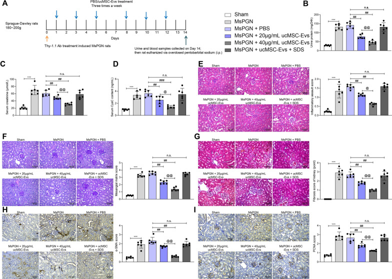
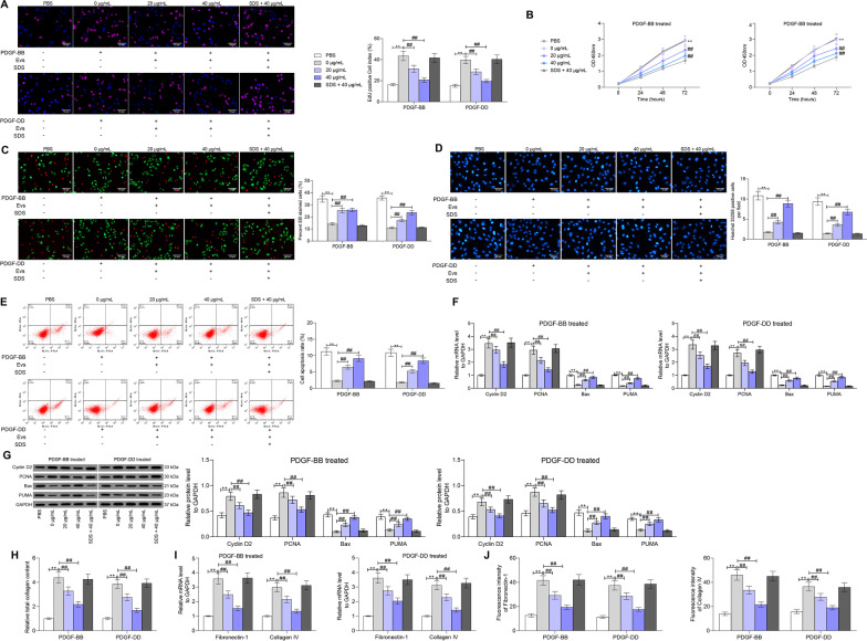
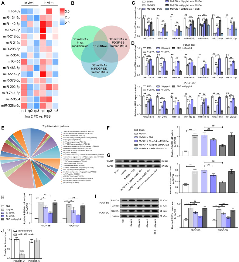
A rat model of MsPGN was induced by anti-Thy-1.1, and rat mesangial cells (rMCs) HBZY-1 were treated with PDGF-BB/DD to mimic MsPGN condition in vitro. Rats and cells were treated with different doses of ucMSC-Evs, and then the pathological changes in renal tissues and proliferation of rMCs were determined. Differentially expressed microRNAs (miRNAs) after Evs treatment were screened by microarray analysis. The interactions among miR-378, PSMD14, and TGFBR1 were analyzed. Gain- and loss-of function studies of miR-378 and PSMD14 were performed to explore their effects on tissue hyperplasia and rMC proliferation and their interactions with the TGF-β1/Smad2/3 signaling pathway.
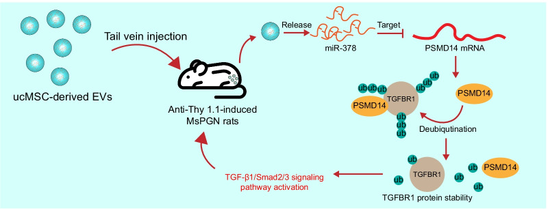
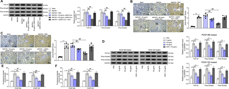
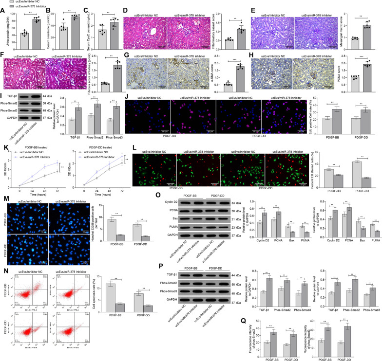
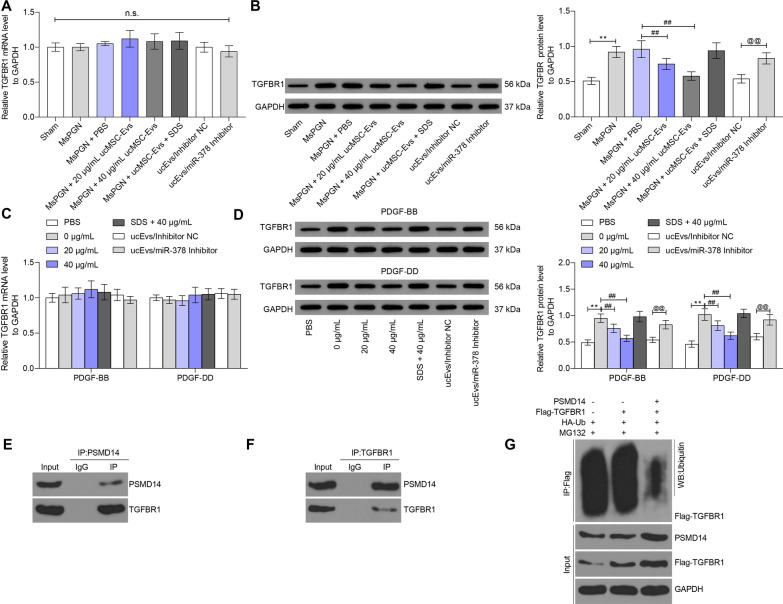
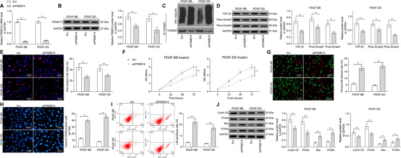
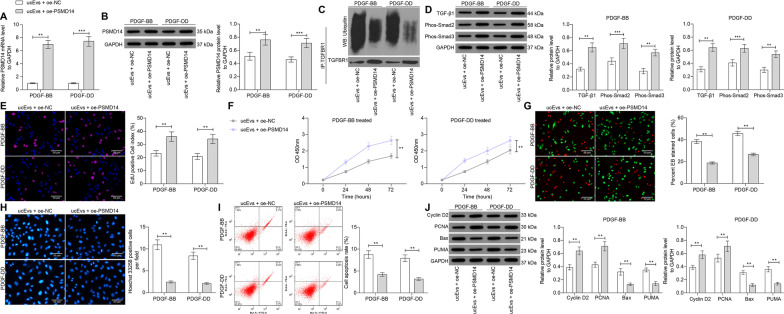
The ucMSC-Evs treatment ameliorated mesangial hyperplasia and fibrosis in rat renal tissues and suppressed the aberrant proliferation of rMCs in a dose-dependent manner. miR-378 was the most upregulated miRNA in tissues and cells after ucMSC-Evs treatment. miR-378 directly targeted PSMD14, and PSMD14 maintained the stability of TGFBR1 through deubiquitination modification, which led to TGF-β1/Smad2/3 activation. Either miR-378 knockdown or PSMD14 overexpression diminished the protective functions of ucMSC-Evs by activating the TGF-β1/Smad2/3 signaling pathway.
UcMSC-Evs ameliorate pathological process in MsPGN through the delivery of miR-378, which suppresses PSMD14-mediated TGFBR1 stability and inactivates the TGF-β1/Smad2/3 signaling pathway to reduce tissue hyperplasia and rMC proliferation. Video abstract.
间充质干细胞(MSCs)及其释放的细胞外囊泡(Evs)已显示出对肾脏疾病的保护作用。本研究旨在研究脐带 MSC 释放的 Evs(ucMSC-Evs)及其在系膜增生性肾小球肾炎(MsPGN)中的作用。
通过抗 Thy-1.1 诱导大鼠 MsPGN 模型,并用 PDGF-BB/DD 处理大鼠系膜细胞(rMCs)HBZY-1 以模拟 MsPGN 条件。用不同剂量的 ucMSC-Evs 处理大鼠和细胞,然后测定肾组织的病理变化和 rMC 的增殖。通过微阵列分析筛选 Evs 处理后差异表达的 microRNAs(miRNAs)。分析 miR-378、PSMD14 和 TGFBR1 之间的相互作用。进行 miR-378 和 PSMD14 的功能获得和功能丧失研究,以探讨它们对组织增生和 rMC 增殖的影响及其与 TGF-β1/Smad2/3 信号通路的相互作用。
ucMSC-Evs 治疗可改善大鼠肾组织系膜增生和纤维化,并呈剂量依赖性抑制 rMC 异常增殖。ucMSC-Evs 处理后组织和细胞中 miR-378 表达上调最明显。miR-378 直接靶向 PSMD14,PSMD14 通过去泛素化修饰维持 TGFBR1 的稳定性,从而激活 TGF-β1/Smad2/3 信号通路。miR-378 敲低或 PSMD14 过表达均通过激活 TGF-β1/Smad2/3 信号通路减弱 ucMSC-Evs 的保护作用。
ucMSC-Evs 通过递送 miR-378 改善 MsPGN 的病理过程,miR-378 抑制 PSMD14 介导的 TGFBR1 稳定性并使 TGF-β1/Smad2/3 信号通路失活,从而减少组织增生和 rMC 增殖。视频摘要。