Vassantachart April, Cao Yufeng, Gribble Michael, Guzman Samuel, Ye Jason C, Hurth Kyle, Mathew Anna, Zada Gabriel, Fan Zhaoyang, Chang Eric L, Yang Wensha
Department of Radiation Oncology, LAC+USC Medical Center, Los Angeles, CA, USA.
Department of Radiation Oncology, Medical Physics Division, Norris Cancer Hospital, Keck School of Medicine, University of Southern California, 1441 Eastlake Ave., Los Angeles, CA, 90033, USA.
Sci Rep. 2022 Mar 9;12(1):3806. doi: 10.1038/s41598-022-07859-0.
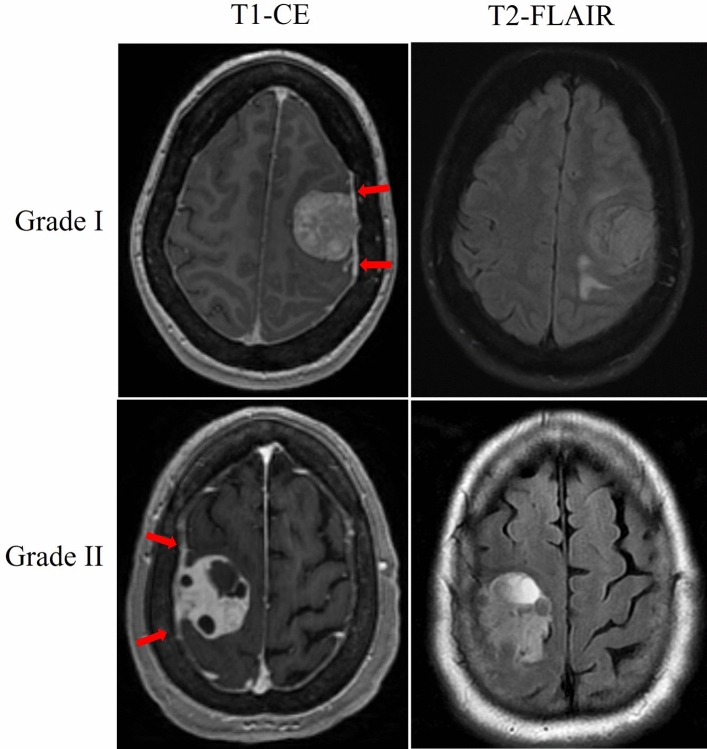
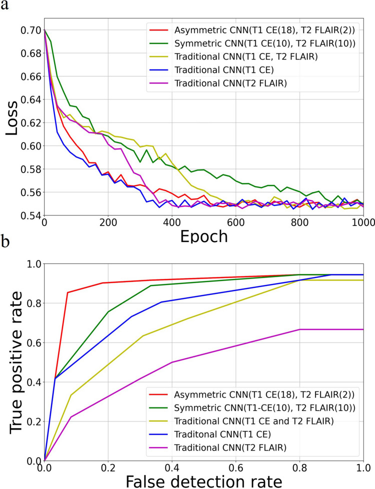
The Grade of meningioma has significant implications for selecting treatment regimens ranging from observation to surgical resection with adjuvant radiation. For most patients, meningiomas are diagnosed radiologically, and Grade is not determined unless a surgical procedure is performed. The goal of this study is to train a novel auto-classification network to determine Grade I and II meningiomas using T1-contrast enhancing (T1-CE) and T2-Fluid attenuated inversion recovery (FLAIR) magnetic resonance (MR) images. Ninety-six consecutive treatment naïve patients with pre-operative T1-CE and T2-FLAIR MR images and subsequent pathologically diagnosed intracranial meningiomas were evaluated. Delineation of meningiomas was completed on both MR images. A novel asymmetric 3D convolutional neural network (CNN) architecture was constructed with two encoding paths based on T1-CE and T2-FLAIR. Each path used the same 3 × 3 × 3 kernel with different filters to weigh the spatial features of each sequence separately. Final model performance was assessed by tenfold cross-validation. Of the 96 patients, 55 (57%) were pathologically classified as Grade I and 41 (43%) as Grade II meningiomas. Optimization of our model led to a filter weighting of 18:2 between the T1-CE and T2-FLAIR MR image paths. 86 (90%) patients were classified correctly, and 10 (10%) were misclassified based on their pre-operative MRs with a model sensitivity of 0.85 and specificity of 0.93. Among the misclassified, 4 were Grade I, and 6 were Grade II. The model is robust to tumor locations and sizes. A novel asymmetric CNN with two differently weighted encoding paths was developed for successful automated meningioma grade classification. Our model outperforms CNN using a single path for single or multimodal MR-based classification.
脑膜瘤的分级对于选择从观察到手术切除并辅以放疗的治疗方案具有重要意义。对于大多数患者而言,脑膜瘤通过影像学诊断,除非进行手术,否则无法确定分级。本研究的目的是训练一种新型自动分类网络,使用T1加权增强(T1-CE)和T2加权液体衰减反转恢复(FLAIR)磁共振(MR)图像来确定I级和II级脑膜瘤。对96例连续的未经治疗且具有术前T1-CE和T2-FLAIR MR图像以及随后经病理诊断为颅内脑膜瘤的患者进行了评估。在两种MR图像上均完成了脑膜瘤的勾勒。构建了一种新型非对称3D卷积神经网络(CNN)架构,该架构基于T1-CE和T2-FLAIR有两条编码路径。每条路径使用相同的3×3×3内核和不同的滤波器,以分别权衡每个序列的空间特征。通过十折交叉验证评估最终模型的性能。在这96例患者中,55例(57%)病理分类为I级脑膜瘤,41例(43%)为II级脑膜瘤。对我们模型的优化导致T1-CE和T2-FLAIR MR图像路径之间的滤波器权重为18:2。基于术前MR,86例(90%)患者分类正确,10例(10%)被错误分类,模型敏感性为0.85,特异性为0.93。在错误分类的患者中,4例为I级,6例为II级。该模型对肿瘤位置和大小具有鲁棒性。开发了一种具有两条权重不同的编码路径的新型非对称CNN,用于成功实现脑膜瘤分级的自动分类。我们的模型在基于单模态或多模态MR的分类中,优于使用单一路径的CNN。