• 文献检索
  • 文档翻译
  • 深度研究
  • 学术资讯
  • Suppr Zotero 插件Zotero 插件
  • 邀请有礼
  • 套餐&价格
  • 历史记录
应用&插件
Suppr Zotero 插件Zotero 插件浏览器插件Mac 客户端Windows 客户端微信小程序
定价
高级版会员购买积分包购买API积分包
服务
文献检索文档翻译深度研究API 文档MCP 服务
关于我们
关于 Suppr公司介绍联系我们用户协议隐私条款
关注我们

Suppr 超能文献

核心技术专利:CN118964589B侵权必究
粤ICP备2023148730 号-1Suppr @ 2026

文献检索

告别复杂PubMed语法,用中文像聊天一样搜索,搜遍4000万医学文献。AI智能推荐,让科研检索更轻松。

立即免费搜索

文件翻译

保留排版,准确专业,支持PDF/Word/PPT等文件格式,支持 12+语言互译。

免费翻译文档

深度研究

AI帮你快速写综述,25分钟生成高质量综述,智能提取关键信息,辅助科研写作。

立即免费体验

多机构脑膜瘤 MRI 数据集,用于自动多序列图像分割。

A multi-institutional meningioma MRI dataset for automated multi-sequence image segmentation.

机构信息

Department of Radiation Oncology, Duke University Medical Center, Durham, NC, USA.

Department of Neurosurgery, Thomas Jefferson University, Philadelphia, PA, USA.

出版信息

Sci Data. 2024 May 15;11(1):496. doi: 10.1038/s41597-024-03350-9.

DOI:10.1038/s41597-024-03350-9
PMID:38750041
原文链接:https://pmc.ncbi.nlm.nih.gov/articles/PMC11096318/
Abstract

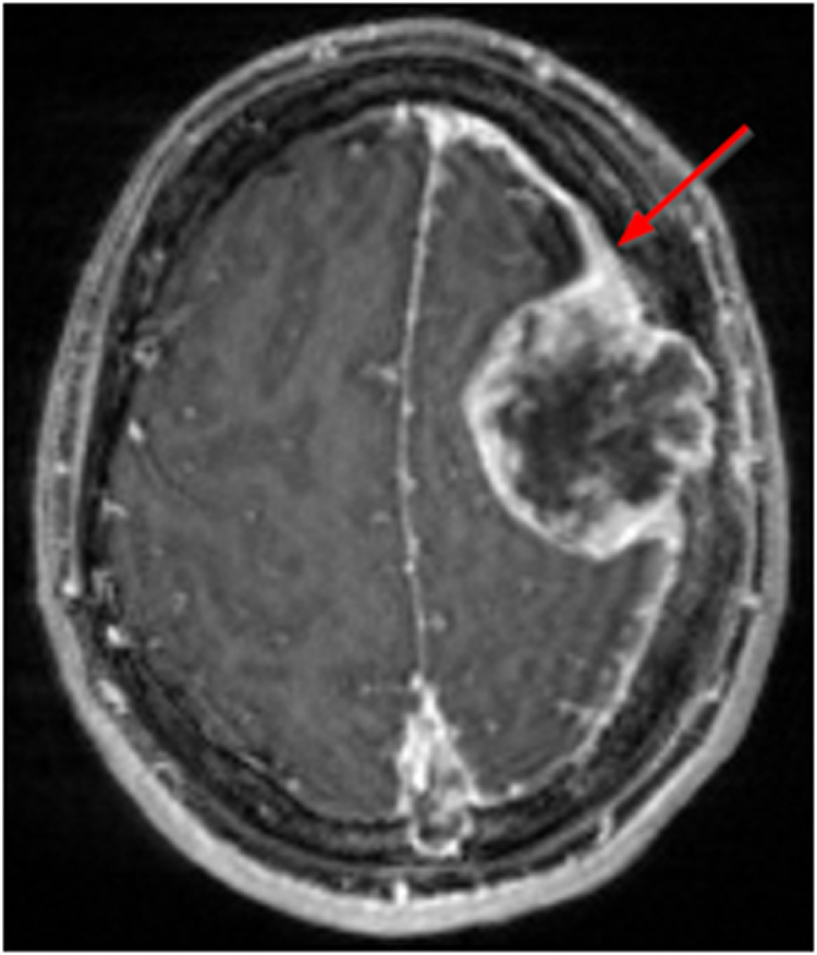
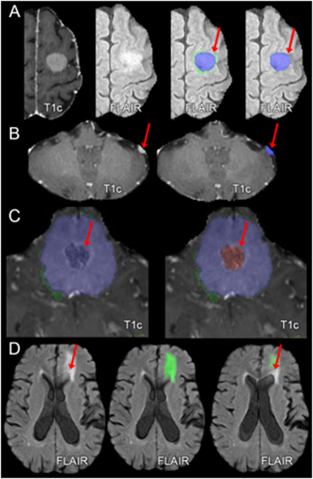
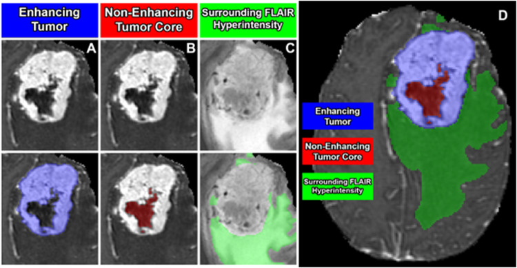
Meningiomas are the most common primary intracranial tumors and can be associated with significant morbidity and mortality. Radiologists, neurosurgeons, neuro-oncologists, and radiation oncologists rely on brain MRI for diagnosis, treatment planning, and longitudinal treatment monitoring. However, automated, objective, and quantitative tools for non-invasive assessment of meningiomas on multi-sequence MR images are not available. Here we present the BraTS Pre-operative Meningioma Dataset, as the largest multi-institutional expert annotated multilabel meningioma multi-sequence MR image dataset to date. This dataset includes 1,141 multi-sequence MR images from six sites, each with four structural MRI sequences (T2-, T2/FLAIR-, pre-contrast T1-, and post-contrast T1-weighted) accompanied by expert manually refined segmentations of three distinct meningioma sub-compartments: enhancing tumor, non-enhancing tumor, and surrounding non-enhancing T2/FLAIR hyperintensity. Basic demographic data are provided including age at time of initial imaging, sex, and CNS WHO grade. The goal of releasing this dataset is to facilitate the development of automated computational methods for meningioma segmentation and expedite their incorporation into clinical practice, ultimately targeting improvement in the care of meningioma patients.

摘要

脑膜瘤是最常见的原发性颅内肿瘤,可导致严重的发病率和死亡率。放射科医生、神经外科医生、神经肿瘤学家和放射肿瘤学家依赖脑部 MRI 进行诊断、治疗计划和纵向治疗监测。然而,目前还没有用于多序列 MR 图像上非侵袭性评估脑膜瘤的自动化、客观和定量工具。在这里,我们介绍了 BraTS 术前脑膜瘤数据集,这是迄今为止最大的多机构专家注释多标签脑膜瘤多序列 MR 图像数据集。该数据集包括来自六个地点的 1141 多序列 MR 图像,每个图像都有四个结构 MRI 序列(T2-、T2/FLAIR-、对比前 T1-和对比后 T1 加权),并附有专家手动细化的三个不同脑膜瘤亚区的分割:增强肿瘤、非增强肿瘤和周围非增强 T2/FLAIR 高信号。提供了基本的人口统计学数据,包括初始成像时的年龄、性别和中枢神经系统世界卫生组织分级。发布此数据集的目的是促进用于脑膜瘤分割的自动化计算方法的开发,并加速其纳入临床实践,最终目标是改善脑膜瘤患者的护理。

https://cdn.ncbi.nlm.nih.gov/pmc/blobs/2d43/11096318/c5c8278193fd/41597_2024_3350_Fig3_HTML.jpg
https://cdn.ncbi.nlm.nih.gov/pmc/blobs/2d43/11096318/d718f1fa5578/41597_2024_3350_Fig1_HTML.jpg
https://cdn.ncbi.nlm.nih.gov/pmc/blobs/2d43/11096318/8a552d2cb2e4/41597_2024_3350_Fig2_HTML.jpg
https://cdn.ncbi.nlm.nih.gov/pmc/blobs/2d43/11096318/c5c8278193fd/41597_2024_3350_Fig3_HTML.jpg
https://cdn.ncbi.nlm.nih.gov/pmc/blobs/2d43/11096318/d718f1fa5578/41597_2024_3350_Fig1_HTML.jpg
https://cdn.ncbi.nlm.nih.gov/pmc/blobs/2d43/11096318/8a552d2cb2e4/41597_2024_3350_Fig2_HTML.jpg
https://cdn.ncbi.nlm.nih.gov/pmc/blobs/2d43/11096318/c5c8278193fd/41597_2024_3350_Fig3_HTML.jpg

相似文献

1
A multi-institutional meningioma MRI dataset for automated multi-sequence image segmentation.多机构脑膜瘤 MRI 数据集,用于自动多序列图像分割。
Sci Data. 2024 May 15;11(1):496. doi: 10.1038/s41597-024-03350-9.
2
The ASNR-MICCAI Brain Tumor Segmentation (BraTS) Challenge 2023: Intracranial Meningioma.2023年ASNR-MICCAI脑肿瘤分割(BraTS)挑战赛:颅内脑膜瘤
ArXiv. 2023 May 12:arXiv:2305.07642v1.
3
Fully automated detection and segmentation of meningiomas using deep learning on routine multiparametric MRI.基于常规多参数 MRI 的深度学习全自动脑膜瘤检测与分割。
Eur Radiol. 2019 Jan;29(1):124-132. doi: 10.1007/s00330-018-5595-8. Epub 2018 Jun 25.
4
Automated Meningioma Segmentation in Multiparametric MRI : Comparable Effectiveness of a Deep Learning Model and Manual Segmentation.多参数磁共振成像中脑膜瘤的自动分割:深度学习模型与手动分割的等效有效性
Clin Neuroradiol. 2021 Jun;31(2):357-366. doi: 10.1007/s00062-020-00884-4. Epub 2020 Feb 14.
5
Fully Automated MRI Segmentation and Volumetric Measurement of Intracranial Meningioma Using Deep Learning.基于深度学习的颅内脑膜瘤全自动 MRI 分割与容量测量。
J Magn Reson Imaging. 2023 Mar;57(3):871-881. doi: 10.1002/jmri.28332. Epub 2022 Jul 1.
6
The Predictive Value of Conventional Magnetic Resonance Imaging Sequences on Operative Findings and Histopathology of Intracranial Meningiomas: A Prospective Study.常规磁共振成像序列对颅内脑膜瘤手术发现和组织病理学的预测价值:一项前瞻性研究。
Neurol India. 2019 Nov-Dec;67(6):1439-1445. doi: 10.4103/0028-3886.273632.
7
Comparison of machine learning classifiers for differentiation of grade 1 from higher gradings in meningioma: A multicenter radiomics study.基于多中心影像组学研究的机器学习分类器在鉴别脑膜瘤 1 级与高级别脑膜瘤中的应用比较。
Magn Reson Imaging. 2019 Nov;63:244-249. doi: 10.1016/j.mri.2019.08.011. Epub 2019 Aug 16.
8
Dynamic susceptibility contrast and dynamic contrast-enhanced MRI characteristics to distinguish microcystic meningiomas from traditional Grade I meningiomas and high-grade gliomas.动态磁敏感对比和动态对比增强 MRI 特征有助于鉴别微囊型脑膜瘤与传统Ⅰ级脑膜瘤和高级别胶质瘤。
J Neurosurg. 2017 Apr;126(4):1220-1226. doi: 10.3171/2016.3.JNS14243. Epub 2016 Jun 10.
9
Accuracy of Radiomics-Based Feature Analysis on Multiparametric Magnetic Resonance Images for Noninvasive Meningioma Grading.基于多参数磁共振成像的放射组学特征分析对脑膜瘤分级的准确性。
World Neurosurg. 2019 Dec;132:e366-e390. doi: 10.1016/j.wneu.2019.08.148. Epub 2019 Aug 30.
10
Deep learning-based automatic segmentation of meningioma from multiparametric MRI for preoperative meningioma differentiation using radiomic features: a multicentre study.基于深度学习的多参数 MRI 脑膜瘤自动分割用于术前脑膜瘤分化的放射组学特征:一项多中心研究。
Eur Radiol. 2022 Oct;32(10):7248-7259. doi: 10.1007/s00330-022-08749-9. Epub 2022 Apr 14.

引用本文的文献

1
Overview of the Head and Neck Tumor Segmentation for Magnetic Resonance Guided Applications (HNTS-MRG) 2024 Challenge.2024年磁共振引导应用头颈肿瘤分割挑战赛(HNTS-MRG)概述。
Head Neck Tumor Segm MR Guid Appl (2024). 2025;15273:1-35. doi: 10.1007/978-3-031-83274-1_1. Epub 2025 Mar 3.
2
Overview of the Head and Neck Tumor Segmentation for Magnetic Resonance Guided Applications (HNTS-MRG) 2024 Challenge.2024年磁共振引导应用头颈肿瘤分割(HNTS-MRG)挑战赛概述
ArXiv. 2024 Nov 28:arXiv:2411.18585v2.
3
A Practical Guide to Manual and Semi-Automated Neurosurgical Brain Lesion Segmentation.

本文引用的文献

1
Federated learning enables big data for rare cancer boundary detection.联邦学习为罕见癌症边界检测提供大数据支持。
Nat Commun. 2022 Dec 5;13(1):7346. doi: 10.1038/s41467-022-33407-5.
2
The federated tumor segmentation (FeTS) tool: an open-source solution to further solid tumor research.联邦肿瘤分割(FeTS)工具:一个开源解决方案,用于进一步的实体瘤研究。
Phys Med Biol. 2022 Oct 12;67(20). doi: 10.1088/1361-6560/ac9449.
3
Automatic differentiation of Grade I and II meningiomas on magnetic resonance image using an asymmetric convolutional neural network.
《手动和半自动神经外科脑病变分割实用指南》
NeuroSci. 2024 Aug 2;5(3):265-275. doi: 10.3390/neurosci5030021. eCollection 2024 Sep.
4
A large scale multi institutional study for radiomics driven machine learning for meningioma grading.大规模多机构研究用于脑膜瘤分级的放射组学驱动的机器学习。
Sci Rep. 2024 Oct 31;14(1):26191. doi: 10.1038/s41598-024-78311-8.
使用非对称卷积神经网络在磁共振图像上对I级和II级脑膜瘤进行自动鉴别。
Sci Rep. 2022 Mar 9;12(1):3806. doi: 10.1038/s41598-022-07859-0.
4
Delphi methodology in healthcare research: How to decide its appropriateness.医疗保健研究中的德尔菲法:如何确定其适用性。
World J Methodol. 2021 Jul 20;11(4):116-129. doi: 10.5662/wjm.v11.i4.116.
5
The 2021 WHO Classification of Tumors of the Central Nervous System: a summary.2021 年世卫组织中枢神经系统肿瘤分类:概述。
Neuro Oncol. 2021 Aug 2;23(8):1231-1251. doi: 10.1093/neuonc/noab106.
6
Meningioma: A Review of Epidemiology, Pathology, Diagnosis, Treatment, and Future Directions.脑膜瘤:流行病学、病理学、诊断、治疗及未来方向综述
Biomedicines. 2021 Mar 21;9(3):319. doi: 10.3390/biomedicines9030319.
7
Meningioma: A Review of Clinicopathological and Molecular Aspects.脑膜瘤:临床病理及分子学方面综述
Front Oncol. 2020 Oct 23;10:579599. doi: 10.3389/fonc.2020.579599. eCollection 2020.
8
Brain extraction on MRI scans in presence of diffuse glioma: Multi-institutional performance evaluation of deep learning methods and robust modality-agnostic training.MRI 扫描中存在弥漫性胶质瘤时的脑提取:深度学习方法的多机构性能评估和稳健的模态不可知训练。
Neuroimage. 2020 Oct 15;220:117081. doi: 10.1016/j.neuroimage.2020.117081. Epub 2020 Jun 27.
9
High-risk Meningioma: Initial Outcomes From NRG Oncology/RTOG 0539.高危脑膜瘤:NRG 肿瘤学/RTOG 0539 的初步结果。
Int J Radiat Oncol Biol Phys. 2020 Mar 15;106(4):790-799. doi: 10.1016/j.ijrobp.2019.11.028. Epub 2019 Nov 29.
10
Trends in intracranial meningioma incidence in the United States, 2004-2015.美国颅内脑膜瘤发病率趋势,2004-2015 年。
Cancer Med. 2019 Oct;8(14):6458-6467. doi: 10.1002/cam4.2516. Epub 2019 Sep 1.