Clem Jones Centre for Ageing Dementia Research, Queensland Brain Institute, The University of Queensland, Brisbane (St Lucia Campus), QLD 4072, Australia.
Cell and Molecular Biology Department, Mental Health Program, QIMR Berghofer Medical Research Institute, Brisbane, QLD 4006, Australia.
Theranostics. 2022 Jan 31;12(5):1952-1970. doi: 10.7150/thno.65539. eCollection 2022.
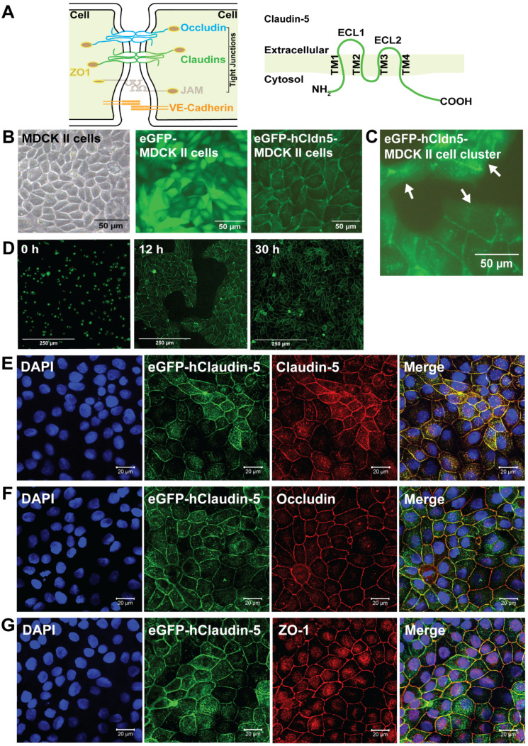
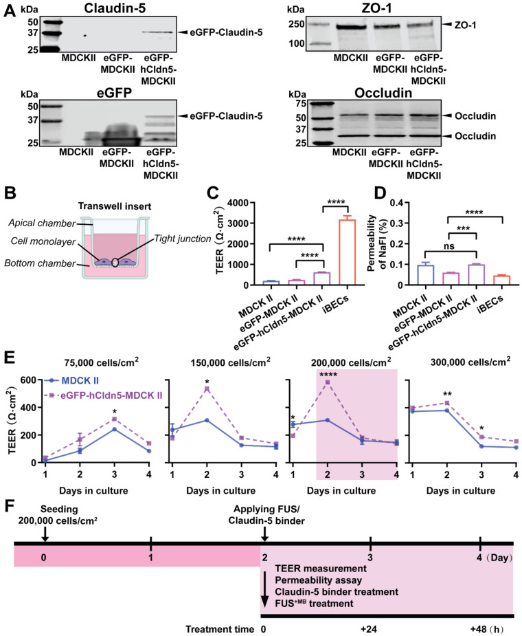
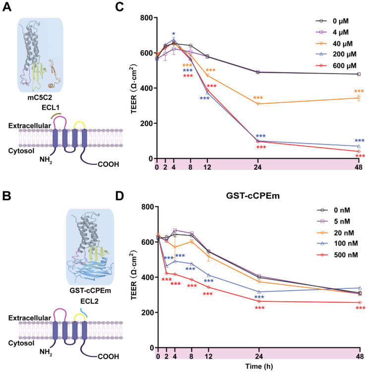
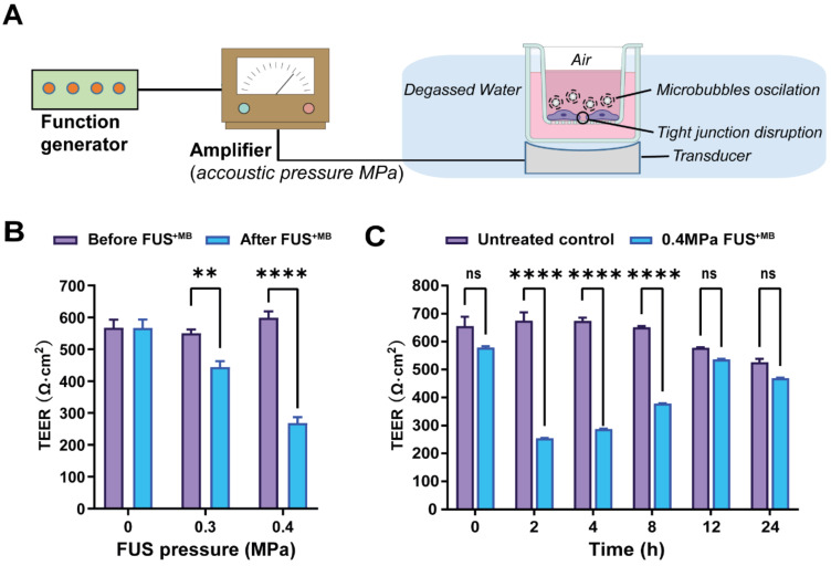
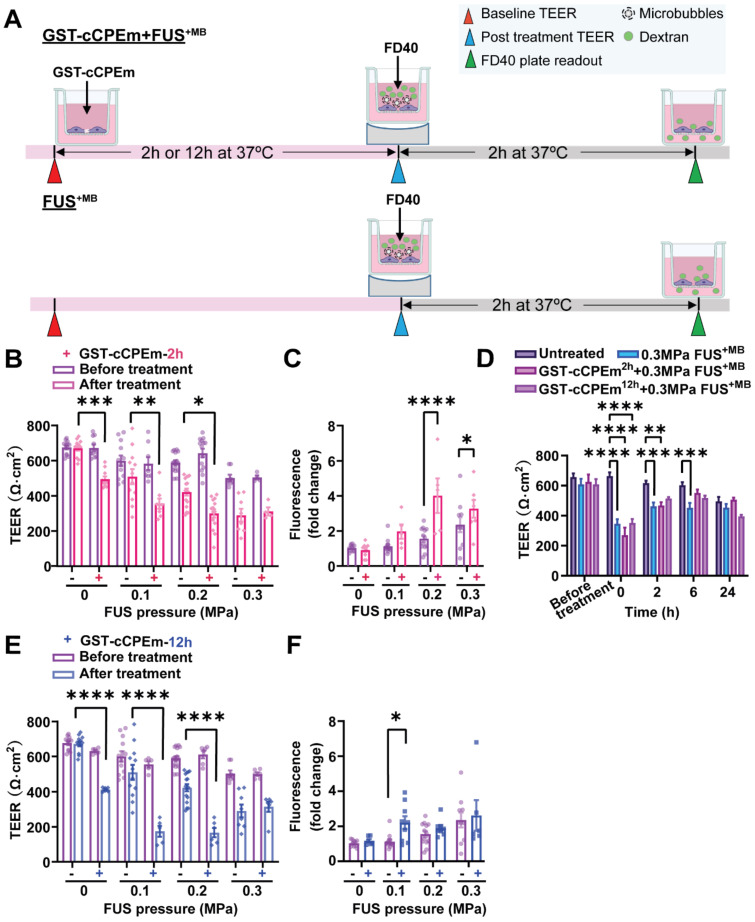
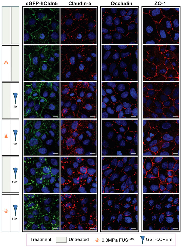
The blood-brain barrier (BBB) while functioning as a gatekeeper of the brain, impedes cerebral drug delivery. An emerging technology to overcome this limitation is focused ultrasound (FUS). When FUS interacts with intravenously injected microbubbles (FUS), the BBB opens, transiently allowing the access of therapeutic agents into the brain. However, the ultrasound parameters need to be tightly tuned: when the acoustic pressure is too low there is no opening, and when it is too high, tissue damage can occur. We therefore asked whether barrier permeability can be increased by combining FUS with a second modality such that in a clinical setting lower acoustic pressures could be used. Given that FUS achieves BBB opening in part by disruption of tight junction (TJ) proteins such as claudin-5 of brain endothelial cells, we generated a stable MDCK (Madin-Darby Canine Kidney) II cell line (eGFP-hCldn5-MDCK II) that expresses fluorescently tagged human claudin-5. Two claudin-5 binders, the peptide mC5C2 and cCPEm (truncated form of an enterotoxin), reported previously to weaken the barrier, were synthesized and assessed for their abilities to enhance the permeability of cellular monolayers. We then performed a comparative analysis of single and combination treatments, measuring transendothelial electrical resistance (TEER) and cargo leakage, combined with confocal image analysis. We successfully generated a novel cell line that formed functional monolayers as validated by an increased TEER reading and a low (< 0.2%) permeability to sodium fluorescein (376 Da). We found that the binders exerted a time- and concentration-dependent effect on barrier opening when incubated over an extended period, whereas FUS caused a rapid opening followed by recovery after 12 hours within the tested pressure range. Importantly, preincubation with cCPEm prior to FUS treatment resulted in greater barrier opening compared to either FUS or cCPEm alone as measured by reduced TEER values and an increased permeability to fluorescently labelled 40 kDa dextran (FD40). The data suggest that pre incubation with clinically suitable binders to TJ proteins may be a general strategy to facilitate safer and more effective ultrasound-mediated BBB opening in cellular and animal systems and potentially also for the treatment of human diseases of the brain.
血脑屏障 (BBB) 作为大脑的守门员,阻碍了脑内药物的输送。一种克服这一限制的新兴技术是聚焦超声 (FUS)。当 FUS 与静脉内注射的微泡 (FUS) 相互作用时,BBB 打开,短暂地允许治疗剂进入大脑。然而,超声参数需要紧密调整:当声压过低时,没有开口,当声压过高时,可能会发生组织损伤。因此,我们询问是否可以通过将 FUS 与第二种模式结合使用来增加屏障通透性,以便在临床环境中可以使用较低的声压。鉴于 FUS 通过破坏脑内皮细胞的紧密连接 (TJ) 蛋白(如 Claudin-5)部分实现 BBB 开放,我们生成了一种稳定的 MDCK(Madin-Darby Canine Kidney)II 细胞系 (eGFP-hCldn5-MDCK II),该细胞系表达荧光标记的人 Claudin-5。两种 Claudin-5 结合物,即先前报道可削弱屏障的肽 mC5C2 和 cCPEm(肠毒素的截断形式),被合成并评估其增强细胞单层通透性的能力。然后,我们进行了单次和联合治疗的比较分析,测量了跨内皮电阻 (TEER) 和货物渗漏,并结合共聚焦图像分析。我们成功地生成了一种新型细胞系,该细胞系形成了功能单层,这通过增加 TEER 读数和对 376 Da 的荧光素钠 (< 0.2%) 的低通透性得到验证。我们发现,当孵育时间延长时,结合物对屏障开放具有时间和浓度依赖性的作用,而 FUS 在测试压力范围内 12 小时内会导致快速开放,随后恢复。重要的是,在用 FUS 处理之前用 cCPEm 预孵育会导致更大的屏障开放,这可以通过降低 TEER 值和增加对荧光标记的 40 kDa 葡聚糖 (FD40) 的通透性来衡量。数据表明,用临床适用的 TJ 蛋白结合物预孵育可能是一种通用策略,可促进细胞和动物系统中更安全、更有效的超声介导的 BBB 开放,并可能也适用于治疗人类脑部疾病。