Imaging and Data Analytics, Clinical Pharmacology & Safety Sciences, R&D, AstraZeneca, Cambridge, UK.
Department of Chemistry, Technical University of Munich, Garching, Germany.
Theranostics. 2022 Feb 14;12(5):2162-2174. doi: 10.7150/thno.68000. eCollection 2022.
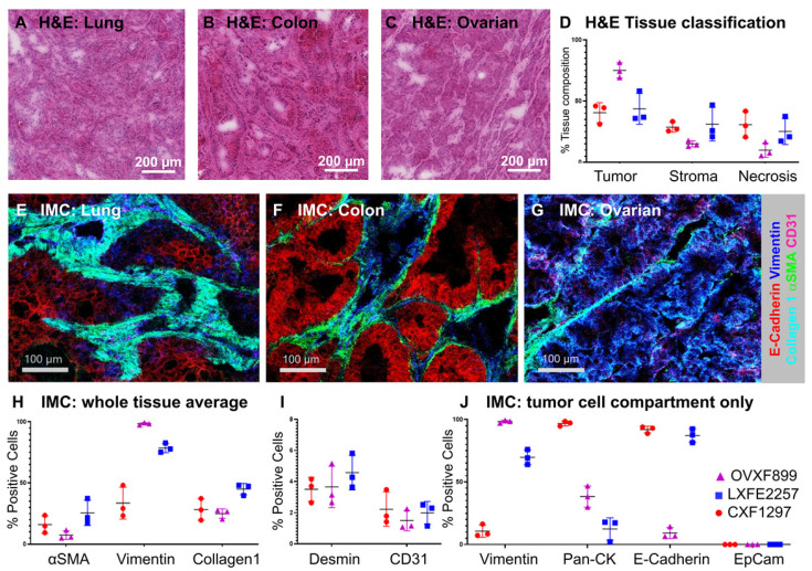
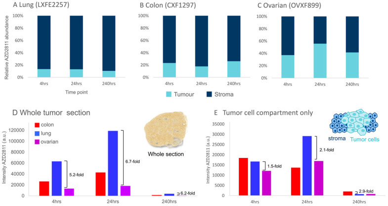
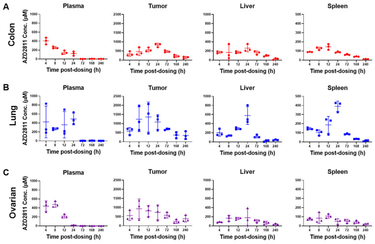
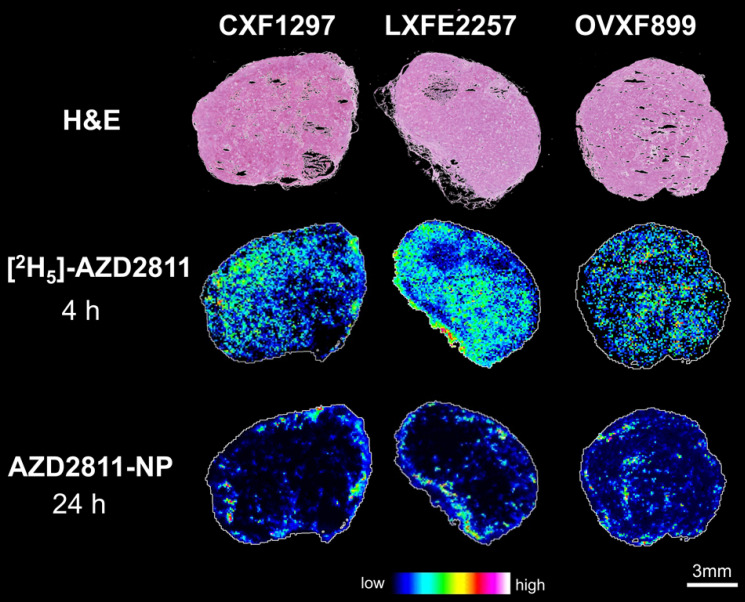
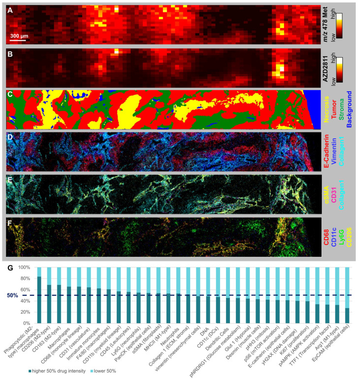
Gaining insight into the heterogeneity of nanoparticle drug distribution within tumors would improve both design and clinical translation of nanomedicines. There is little data showing the spatio-temporal behavior of nanomedicines in tissues as current methods are not able to provide a comprehensive view of the nanomedicine distribution, released drug or its effects in the context of a complex tissue microenvironment. A new experimental approach which integrates the molecular imaging and bioanalytical technologies MSI and IMC was developed to determine the biodistribution of total drug and drug metabolite delivered via PLA-PEG nanoparticles and to overlay this with imaging of the nanomedicine in the context of detailed tumor microenvironment markers. This was used to assess the nanomedicine AZD2811 in animals bearing three different pre-clinical PDX tumors. This new approach delivered new insights into the nanoparticle/drug biodistribution. Mass spectrometry imaging was able to differentiate the tumor distribution of co-dosed deuterated non-nanoparticle-formulated free drug alongside the nanoparticle-formulated drug by directly visualizing both delivery approaches within the same animal or tissue. While the IV delivered free drug was uniformly distributed, the nanomedicine delivered drug was heterogeneous. By staining for multiple biomarkers of the tumor microenvironment on the same tumor sections using imaging mass cytometry, co-registering and integrating data from both imaging modalities it was possible to determine the features in regions with highest nanomedicine distribution. Nanomedicine delivered drug was associated with regions higher in macrophages, as well as more stromal regions of the tumor. Such a comparison of complementary molecular data allows delineation of drug abundance in individual cell types and in stroma. This multi-modal imaging solution offers researchers a better understanding of drug and nanocarrier distribution in complex tissues and enables data-driven drug carrier design.
深入了解纳米药物在肿瘤内的异质性分布将提高纳米药物的设计和临床转化。目前的方法无法提供纳米药物在复杂组织微环境中的分布、释放药物或其作用的全面视图,因此几乎没有数据显示纳米药物在组织中的时空行为。一种新的实验方法,将分子成像和生物分析技术 MSI 和 IMC 相结合,用于确定通过 PLA-PEG 纳米粒子递送的总药物和药物代谢物的生物分布,并将其与纳米药物在详细肿瘤微环境标志物背景下的成像叠加。这用于评估在携带三种不同临床前 PDX 肿瘤的动物中的纳米药物 AZD2811。这种新方法深入了解了纳米颗粒/药物的生物分布。质谱成像能够通过直接在同一动物或组织内可视化两种给药途径,区分共给药氘标记的非纳米颗粒制剂游离药物与纳米颗粒制剂药物的肿瘤分布。虽然静脉内递送的游离药物均匀分布,但纳米药物递送的药物则不均匀。通过在同一肿瘤切片上使用成像质谱细胞术对肿瘤微环境的多个生物标志物进行染色,对来自两种成像模式的数据进行配准和整合,有可能确定具有最高纳米药物分布的区域的特征。纳米药物递送的药物与巨噬细胞含量较高的区域以及肿瘤的基质区域有关。这种对互补分子数据的比较可以在单个细胞类型和基质中划定药物丰度。这种多模态成像解决方案为研究人员提供了对复杂组织中药物和纳米载体分布的更好理解,并能够实现基于数据的药物载体设计。